. 2025 Aug 27:8:45.
doi: 10.20517/cdr.2025.103. eCollection 2025.

Cryptotanshinone differentially induces cell death in ATP6V0D1-deficient pancreatic cancer cells

Affiliations

Cryptotanshinone differentially induces cell death in ATP6V0D1-deficient pancreatic cancer cells

Fangquan Chen et al. Cancer Drug Resist. .

Abstract

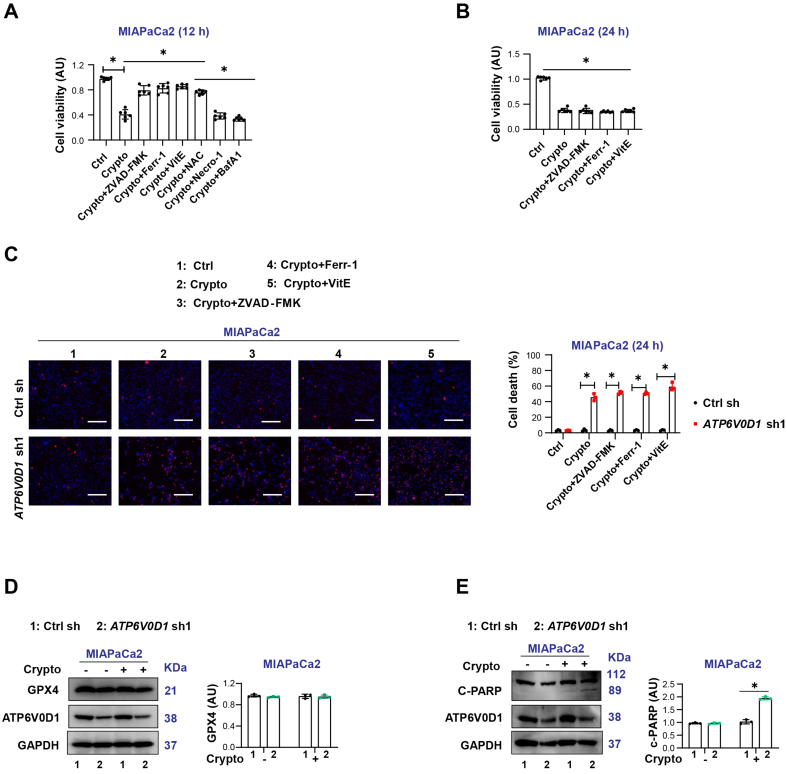
Aim: Dysregulation of tumor-suppressive pathways can lead to constitutive activation of multiple oncogenic signaling cascades. Such overactivation makes cancer cells highly dependent on these pathways, creating potential therapeutic vulnerabilities. Based on our previous findings and current data, genetic knockout of ATPase H+ transporting V0 subunit D1 (ATP6V0D1) - a key mediator of alkaliptosis - induces hyperactivation of oncogenic pathways, including signal transducer and activator of transcription 3 (STAT3)-mediated lysosomal pH regulation and AKT serine/threonine kinase (AKT) signaling. It also alters cellular responses to cryptotanshinone therapy. This study aimed to investigate how ATP6V0D1 deficiency reshapes oncogenic signaling networks and cellular heterogeneity in pancreatic ductal adenocarcinoma (PDAC), while evaluating therapeutic strategies that exploit alkaliptosis-related vulnerabilities. Methods: ATP6V0D1-deficient SW1990 and MIAPaCa2 cells were generated via gene knockdown. Cell viability and death following various treatments were assessed using CCK-8 and propidium iodide assays. Transcriptomic analysis was conducted to identify feedback signaling pathways, while Western blotting was used to measure expression of signaling proteins. Macropinocytosis was evaluated by TRITC-dextran uptake. Additionally, The Cancer Dependency Map (DepMap) database was analyzed to explore background differences between SW1990 and MIAPaCa2 cells. Results: ATP6V0D1 deletion led to overactivation of STAT3-mediated lysosomal pH regulation and AKT signaling; inhibition of these pathways restored alkaliptosis. Notably, cryptotanshinone selectively induced cell death in ATP6V0D1-deficient MIAPaCa2 cells but not SW1990 cells. Resistance in SW1990 cells was mediated by FGFR2 upregulation, which was reversed upon FGFR2 inhibition. Conclusion: ATP6V0D1 deficiency drives PDAC progression via dual mechanisms: compensatory oncogenic signaling (STAT3/AKT) and FGFR2-mediated cellular heterogeneity. While targeting these pathways may offer therapeutic potential, tumor heterogeneity remains a major clinical challenge.

Keywords: ATP6V0D1; cell death; cryptotanshinone; tumor heterogeneity.


Conflict of interest statement

All authors declared that there are no conflicts of interest.

Figures

Figure 1
Figure 1
Macropinocytosis is not responsible for cryptotanshinone-induced differential sensitization of PDAC cells. (A) CCK-8 assay assessing cell viability after treatment with JTC801, cryptotanshinone (Crypto), or their combination (Drug concentrations: JTC801, 3.5 μM; Crypto, 5 μM; treatment time: 24 h); (B) Cell death assay evaluating the proportion of dead cells under the indicated treatments (Same concentrations and treatment time as A); (C) Western blot analysis of protein expression in the indicated groups (Drug concentration: JTC801, 3.5 μM; treatment time: 24 h); (D) TRITC-dextran uptake to assess macropinocytosis in the indicated cell lines (Drug concentration: JTC801, 3.5 μM; treatment time: 24 h). Scale bar = 50 µm; (E) CCK-8 assay assessing cell viability after treatment with JTC801, Crypto, or Crypto combined with 5% BSA; (F) CCK-8 assay evaluating cell viability after treatment with JTC801, Crypto, or Crypto combined with EIPA (Drug concentrations: JTC801, 3.5 μM; Crypto, 5 μM; EIPA, 5 μM; treatment time: 24 h); (G and H) CCK-8 assay assessing the effect of LY2090314 combined with JTC801 (G) or Crypto (H) on cell viability (Drug concentrations: JTC801, 3.5 μM; Crypto, 5 μM; LY2090314, 5 μM; treatment time: 24 h); (I) CCK-8 assay analyzing MIAPaCa2 cell viability treated with Crypto (5 μM) with ATP6V0D1 knockout alone or simultaneous GSK3B knockout. Data (A and B, D-I) are presented as mean ± SD from ≥ 3 biologically independent samples (A, n = 6; B = 4; D, n = 3; E-I, n = 6). Statistical significance was determined using two-way ANOVA with Tukey’s multiple comparisons test. Western blot data (C) represent three independent experiments. Source data are provided. *P < 0.05; ns: not significant. PDAC: Pancreatic ductal adenocarcinoma; ATP6V0D1: ATPase H+ transporting V0 subunit D1; SD: standard deviation; ANOVA: analysis of variance.
Figure 2
Figure 2
PI3K-AKT signaling induces alkaliptosis resistance but does not affect cryptotanshinone sensitivity. (A) KEGG pathway analysis of DEGs based on mRNA transcriptomics; (B) Western blot analysis of indicated proteins in the indicated groups (Drug concentration: JTC801, 3.5 μM; treatment time: 24 h); (C) CCK-8 assay measuring cell viability after treatment with JTC801 or Crypto combined with MK-2206 (Drug concentrations: JTC801, 3.5 μM; MK-2206, 2 μM; treatment time: 24 h); (D) Cell death assay showing the proportion of dead cells following treatment with JTC801, MK-2206, or their combination (same concentrations and time as above). Data (C and D) are shown as mean ± SD from three or more biologically independent samples (C, n = 6; D, n = 3). Statistical significance was determined using two-way ANOVA with Tukey’s multiple comparisons test. Western blots (B) represent three independent experiments. Source data are provided. *P < 0.05 was considered statistically significant; ns: not significant. PI3K: Phosphatidylinositol-4,5-bisphosphate 3-kinase; AKT: AKT serine/threonine kinase; KEGG: Kyoto Encyclopedia of Genes and Genomes; DEGs: differentially expressed genes; SD: standard deviation; ANOVA: analysis of variance.
Figure 3
Figure 3
Differential FGFR2 expression partially determines cryptotanshinone sensitivity in ATP6V0D1-deficient cells. (A and B) Western blot analysis of indicated proteins after treatment (Drug concentrations: JTC801, 3.5 μM; Crypto, 5 μM; LY2874455, 450 nM; treatment time: 24 h); (C) CCK-8 assay of cell viability after JTC801, LY2874455, or combination treatment (same concentrations and time as above); (D) Cell death assay showing mortality after JTC801, LY2874455, or combination treatment; (E) CCK-8 assay evaluating cell viability of SW1990 cells treated with Crypto (5 μM) after ATP6V0D1 knockout or combined ATP6V0D1 and FGFR2 knockout; (F) Cell death assay for the indicated groups (Drug concentration: Crypto, 5 μM; treatment time: 24 h). Data (C-E) are shown as mean ± SD from three or more biologically independent samples (C and D, n = 3; E, n = 6; F, n = 3). Statistical significance was performed using two-way ANOVA with Tukey’s multiple comparisons test. Western blots (A and B) represent three independent experiments. Source data are provided. *P < 0.05 was considered statistically significant; ns: not significant. FGFR2: Fibroblast growth factor receptor 2; ATP6V0D1: ATPase H+ transporting V0 subunit D1; SD: standard deviation; ANOVA: analysis of variance.
Figure 4
Figure 4
Differential expression of FGFR2 partially determines cryptotanshinone sensitivity in ATP6V0D1-deficient cells. (A) CCK-8 assay showing cell viability after treatment with Crypto alone or in combination with various cell death inhibitors (Drug concentrations: Crypto, 5 μM; ZVAD-FMK, 50 μM; Ferr-1, 500 nM; VitE, 5 mM; NAC, 5 mM; Necro-1, 500 nM; Baf A1, 500 nM; treatment time: 12 h); (B) CCK-8 assay showing cell viability after treatment with Crypto alone or in combination with inhibitors (Drug concentrations: Crypto, 5 μM; ZVAD-FMK, 50 μM; Ferr-1, 500 nM; VitE, 5 mM; treatment time: 24 h); (C) Cell death assay showing the proportion of dead cells following treatment with Crypto alone or in combination with inhibitors (Drug concentrations: Crypto, 5 μM; ZVAD-FMK, 50 μM; Ferr-1, 500 Nm, VitE, 5 mM; treatment time: 24 h). Scale bar = 200 µm; (D and E) Western blot analysis showing protein expression in the indicated groups (Drug concentration: Crypto, 5 μM; treatment time: 24 h). Data (A-C) are shown as mean ± SD from at least three biologically independent samples (A and B, n = 6; C, n = 3). Statistical significance was analyzed using two-way ANOVA with Tukey’s multiple comparisons test. Western blots (D and E) represent three independent experiments. Source data are provided. *P < 0.05 was considered statistically significant; ns: not significant. FGFR2: Fibroblast growth factor receptor 2; ATP6V0D1: ATPase H+ transporting V0 subunit D1; SD: standard deviation; ANOVA: analysis of variance.

References

    1. Laplane L, Maley CC. The evolutionary theory of cancer: challenges and potential solutions. Nat Rev Cancer. 2024;24:718–33. doi: 10.1038/s41568-024-00734-2. - DOI - PMC - PubMed
    1. Lee HJ, Zhuang G, Cao Y, Du P, Kim HJ, Settleman J. Drug resistance via feedback activation of Stat3 in oncogene-addicted cancer cells. Cancer Cell. 2014;26:207–21. doi: 10.1016/j.ccr.2014.05.019. - DOI - PubMed
    1. Zeng H, Qu J, Jin N, et al. Feedback activation of leukemia inhibitory factor receptor limits response to histone deacetylase inhibitors in breast cancer. Cancer Cell. 2016;30:459–73. doi: 10.1016/j.ccell.2016.08.001. - DOI - PubMed
    1. Wu Q, Zhen Y, Shi L, et al. EGFR inhibition potentiates FGFR inhibitor therapy and overcomes resistance in FGFR2 fusion-positive cholangiocarcinoma. Cancer Discov. 2022;12:1378–95. doi: 10.1158/2159-8290.CD-21-1168. - DOI - PMC - PubMed
    1. Zhao M, Luo R, Liu Y, et al. miR-3188 regulates nasopharyngeal carcinoma proliferation and chemosensitivity through a FOXO1-modulated positive feedback loop with mTOR-p-PI3K/AKT-c-JUN. Nat Commun. 2016;7:11309. doi: 10.1038/ncomms11309. - DOI - PMC - PubMed

LinkOut - more resources