. 2025 Sep 11:16:1623153.
doi: 10.3389/fphar.2025.1623153. eCollection 2025.

Dehydrozaluzanin C inhibits colon cancer cell proliferation, apoptosis and cycle arrest through peroxisome proliferator-activated receptor γ (PPARγ) activation

Affiliations

Dehydrozaluzanin C inhibits colon cancer cell proliferation, apoptosis and cycle arrest through peroxisome proliferator-activated receptor γ (PPARγ) activation

Shan-Shan Li et al. Front Pharmacol. .

Abstract

Dehydrozaluzanin C (DC) is a sesquiterpene lactone isolated from Asteraceae plant Ainsliaea macrocephala. To investigate the antitumor effects of DC and possible molecular mechanisms for treating cancer. The antitumor effect of DC was studied using HT-29 and HCT-116 human colon tumor cell lines and Balb/c nude mice models. The anti-proliferative, proapoptotic effects, and cycle arrest of DC were observed by cell viability, colony formation, apoptosis, and cycle assays. The changes of protein expression level were examined by Western blot analysis. The transcription activity of PPARγ was determined by Luciferase reporter assay. The role of PPARγ activation in the antitumor activity of DC was verified using PPARγ antagonist GW9662 and si-PPARγ HT-29 cells. DC treatment significantly decreased colon tumor cell viability, cell clone number, and increased apoptosis rate and arrested cell cycle at S phase. Furthermore, DC treatment significantly decreased Bcl-2, CDK2, and cyclin A2 protein levels while increasing the expression of cleaved caspase 3 and Bax in HT-29 and HCT-116 cells. Further investigations indicated that cell survival, induction of apoptosis, and cycle arrest by DC could be significantly reversed following treatment with the PPARγ antagonist GW9662 or in si-PPARγ cells. In vivo, DC treatment significantly decreased the weight and volume of xenograft tumor tissues in mice and apoptosis-related protein levels. The results suggest that DC effectively inhibits colon tumor cell proliferation, clone formation, apoptosis, and cell cycle arrest through PPARγ activation. These results support the potential of DC as an anti-tumor lead compound for further investigation.

Keywords: Dehydrozaluzanin C; PPARγ activation; apoptosis induction; colon cancer; cycle arrest.


Conflict of interest statement

The authors declare that the research was conducted in the absence of any commercial or financial relationships that could be construed as a potential conflict of interest.

Figures
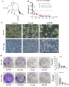
FIGURE 1
FIGURE 1
DC suppressed cell proliferation and colony formation in colon cancer cells. (A) The chemical structure of DC. (B) HT-29, HCT-116 and NCM460 cells were treated at the indicated concentration of DC for 24 h, and cell viability was measured by CCK-8 assay (n = 6). (C) Typical morphological change by DC treatment in HT-29 and HCT-116 cells observed under an inverted light Microscope (Bar = 200 μm). The white arrows indicated that the cells are solidified or fragmented into several granules. (D) The cells were treated by the indicated concentration of DC and stained with 0.5% crystal violet, and photographed with a digital camera. Quantitative analysis of colony formation of HT-29 and HCT-116 cells, respectively (n = 3). Significance was determined by the one-way ANOVA (*** P < 0.001, ** P < 0.01, * P < 0.05 vs. control).
FIGURE 2
FIGURE 2
DC induced cell apoptosis and related-protein expression in colon cancer cells. (A,B) HT-29 and HCT-116 cells were treated with the indicated concentrations of DC for 24 h. The captured cells were fixed and stained by Annexin-V/FITC and PI to analyze the cell apoptotic rates using a flow cytometer. (C,D) The expression of cleaved caspase 3, Bcl-2, and Bax in DC-treated cells for 24 h by Western blotting. Data are expressed as the mean ± S.D. of three independent experiments. Significance was determined by the one-way ANOVA (*** P < 0.001, ** P < 0.01, * P < 0.05 vs. control).
FIGURE 3
FIGURE 3
DC treatment arrested cell cycle at the S phase and regulated the expression of related proteins in colon cancer cells. (A) HT-29 and HCT-116 cells were treated with DC for 24 h. The cells were fixed and stained by PI to analyze the cell cycle distribution using a flow cytometer. (B) Quantification of the cell cycle distribution of HT-29 and HCT-116 cells. (C,D) The expression of CDK2 and Cyclin A2 followed treatment with DC for 24 h by Western blotting. Data are expressed as the mean ± S.D. of three independent experiments. Significance was determined by the one-way ANOVA (*** P < 0.001, ** P < 0.01, * P < 0.05 vs. control).
FIGURE 4
FIGURE 4
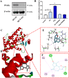
(A) The protein expression of PPARγ in HT-29 and HCT-116 cells. (B) DC increases the transcriptional activity of PPARγ (*** P < 0.001, ** P < 0.01). (C–F) The 3D putative binding modes of DC with the ligand-binding domain (LBD) of PPARγ. (C,D) DC interacts with the key amino acid residues in PPARγ binding pocket (−7.9 kcal/mol). (E,F) DC interact with the Cys285, Leu330, Arg288, Leu333, Ser342, and Met364 in PPARγ binding pocket.
FIGURE 5
FIGURE 5
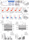
The effect of PPARγ antagonist (GW9662) for DC-mediated antiproliferation, cell colony formation, apoptosis induction and cell cycle arrest in HT-29 cells. HT-29 cells were co-treated with GW9662 and DC for 24 h (A,B) The anti-growth effect of DC was examined by CCK-8 and IC50 value was calculated in HT-29 cells. (C,D) Representative images of colony formation and quantitative analysis of colony-forming ability in HT-29 cells co-treated with GW9662 and DC. (E,F) The apoptosis of HT-29 cells co-treated with GW9662 and DC was determined by using annexin V/PI staining. Quantitative analysis of apoptotic effects in HT-29 cells (n = 3). (G,H) HT-29 cells were stained with PI for the detection of cell cycle distribution. The percentage of cells in each cycling phase is represented by the quantitative analysis (n = 3). (I,J) The protein levels of cleaved caspase-3, Bax, Bcl-2, CDK2 and Cyclin A2 were determined by Western blotting in HT-29 cells. Data are expressed as the mean ± S.D. of three independent experiments. Significance was determined by the one-way ANOVA (*** P < 0.001, ** P < 0.01, * P < 0.05 vs. control).
FIGURE 6
FIGURE 6
The effect of PPARγ knockdown for DC-mediated anti-proliferation, cell colony formation, apoptosis induction and cell cycle arrest in HT-29 cells. The si-NC or si-PPARγ HT-29 cells were treated with DC for 24 h (A,B) The relative expression of PPARγ in si-NC or si-PPARγ HT-29 cells were examined using WB. (C) The antiproliferation effect of DC was examined in PPARγ knockdown HT-29 cells. (D,E) The apoptosis induction of DC was examined by flow cytometry and cell apoptosis rate was calculated. (F,G) The cell cycle distribution was detected by flow cytometry, and the percentage of cells in each cycling phase was represented in the knockdown cells. (H,I) The protein level of cleaved caspase-3, Bax, Bcl-2, CDK2 and Cyclin A2 were determined by Western blotting in PPARγ knockdown HT-29 cells. Data are expressed as the mean ± S.D. of three independent experiments. Significance was determined by the one-way ANOVA (*** P < 0.001, ** P < 0.01, * P < 0.05 vs. control).
FIGURE 7
FIGURE 7
DC suppressed tumor growth in HT-29 cell xenograft mice. HT-29 cells were subcutaneously injected into the right flanks of nude mice, and saline or DC was administrated intraperitoneally daily for 14 days. (A) Time course of body weight (n = 6). (B) Time course of tumor growth progression (n = 6). (C) Tumor weight measured at the end of the experiment (n = 6). (D) Images of the excised tumors at the end of the experiment. (E) Tumor sections were subjected to HE, tunel and immunohistochemistry staining for Ki67. (F,G) The cell number of tunel and ki67 were quantitatively analyzed. (H) The relative protein expression levels of cleaved-caspase 3, Bcl-2, Bax compared to β-actin in the tumor tissues. (I) Detection of apoptosis related proteins by Western blot analysis. (J) Hearts, livers, spleens, lungs, and kidneys were stained with H&E. Data are expressed as the mean ± S.D. of three independent experiments. Significance was determined by the one-way ANOVA (***P < 0.001, **P < 0.01, *P < 0.05 vs. control).

References

    1. Anghel S. I., Wahli W. (2007). Fat poetry: a kingdom for PPAR gamma. Cell Res. 17 (6), 486–511. 10.1038/cr.2007.48 - DOI - PubMed
    1. Atanasov A. G., Zotchev S. B., Dirsch V. M., Orhan I. E., Banach M., Rollinger J. M., et al. (2021). Natural products in drug discovery: advances and opportunities. Nat. Rev. Drug Discov. 20 (3), 200–216. 10.1038/s41573-020-00114-z - DOI - PMC - PubMed
    1. Bray F., Ferlay J., Soerjomataram I., Siegel R. L., Torre L. A., Jemal A. (2018). Global cancer statistics 2018: GLOBOCAN estimates of incidence and mortality worldwide for 36 cancers in 185 countries. CA A Cancer J. Clin. 68 (6), 394–424. 10.3322/caac.21492 - DOI - PubMed
    1. Chen X.-X., Khyeam S., Zhang Z.-J., Zhang K.Y.-B. (2022). Granatin B and punicalagin from Chinese herbal medicine pomegranate peels elicit reactive oxygen species-mediated apoptosis and cell cycle arrest in colorectal cancer cells. Phytomedicine 97, 153923. 10.1016/j.phymed.2022.153923 - DOI - PubMed
    1. Dhyani P., Quispe C., Sharma E., Bahukhandi A., Sati P., Attri D. C., et al. (2022). Anticancer potential of alkaloids: a key emphasis to colchicine, vinblastine, vincristine, vindesine, vinorelbine and vincamine. Cancer Cell Int. 22 (1), 206. 10.1186/s12935-022-02624-9 - DOI - PMC - PubMed

LinkOut - more resources