. 2025 Sep 29;22(1):217.
doi: 10.1186/s12974-025-03554-9.

Cholecystokinin ameliorates cognitive impairment via inhibiting microglia phagocytosis of excitatory synapses in sepsis-associated encephalopathy mice

Affiliations

Cholecystokinin ameliorates cognitive impairment via inhibiting microglia phagocytosis of excitatory synapses in sepsis-associated encephalopathy mice

Lei Chen et al. J Neuroinflammation. .

Abstract

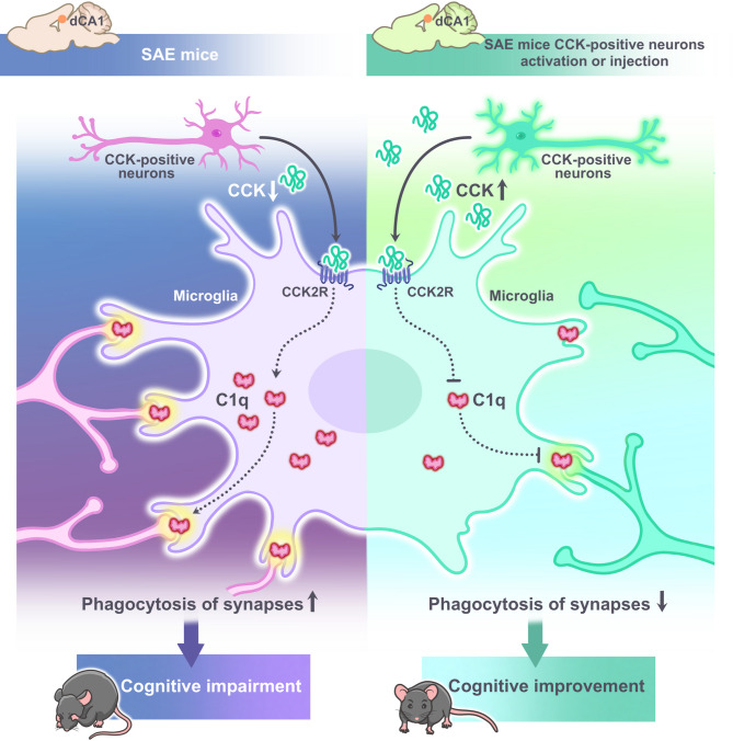
Background: Sepsis-associated encephalopathy (SAE) is characterised by cognitive impairment and is a common complication in patients with sepsis. Microglia are involved in various cognitive impairment-related diseases through phagocytic synapses. Cholecystokinin (CCK), an abundant neuropeptide in the brain, is closely related to cognitive function. However, the role of CCK in SAE and the relationship between CCK and microglial phagocytosis of synapses are unknown.

Methods: Lipopolysaccharide (LPS) was used to construct SAE models in 3-month-old male mice and BV2 microglial cells. To investigate the effects of CCK on cognitive impairment in SAE model mice, we used exogenous CCK injection into the dorsal hippocampal CA1 region or the chemogenetic activation of CCK-positive neurons to promote endogenous CCK release. Morris water maze and fear conditioning test were used to assess cognitive function in mice. RNA sequencing was performed to explore the potential signalling pathways involved in CCK-induced neuroprotection. Western blot and immunofluorescence were used to assess the effects of CCK on microglial phagocytosis of synapses, neurotoxic astrocytes, and excitatory synapses. Whole-cell recording was used to determine excitatory synaptic transmission.

Results: LPS successfully established in vivo and in vitro models of SAE. Both exogenous CCK injection and activation of CCK-positive neurons in hippocampal CA1 region attenuated cognitive impairment in SAE mice. Mechanistically, CCK significantly alleviated excitatory synaptic plasticity damage via inhibiting complement 1q (C1q)-mediated microglial phagocytosis of synapses and neurotoxic astrocyte polarisation. Moreover, in vitro SAE model of BV2 cells demonstrated that CCK exerts neuroprotective effects through microglial CCK2-type receptor.

Conclusions: CCK may alleviate cognitive impairment by inhibiting microglia C1q-mediated phagocytosis of excitatory synapses, suggesting that both CCK drugs and specific activation of CCK-positive neurons are potential treatments for SAE.

Keywords: Cholecystokinin; Complement 1q; Excitatory synapse; Microglia; Sepsis-associated encephalopathy.


Conflict of interest statement

Declarations. Ethics approval and consent to participate: All animal care and experimental procedures were approved by the animal ethics committee of Peking University Health Science Center (ethics number: LA2021534). Consent for publication: Not applicable. Competing interests: The authors declare no competing interests.

Figures

Fig. 1
Fig. 1
Cognitive dysfunction is accompanied by reduced hippocampal cholecystokinin (CCK) and excitatory synapses in sepsis-associated encephalopathy (SAE) model mice. (A) Experimental timeline. ELISA was performed to detect the serum levels of TNF-α (B), IL-1β (C), IL-6 (D), IL-10 (E) and MCP-1 (F), and hippocampal CCK concentration (P). (G) Daily sepsis scores of control mice and mice after lipopolysaccharide (LPS) injection. (H) Daily body weights of control mice and mice after LPS injection. The percentages of freezing time in the (I) training phase, (J) context test, and (K) tone test were recorded on day 1 before and day 3 after the LPS injection. (L) Swimming speed and (M) latency to the platform were recorded in the training and place navigation tests. (N) The number of platform crossings and (O) time spent in the target quadrant were recorded in the Morris Water Maze (MWM) spatial probe test on day 7 after LPS injection. (Q, R) Protein levels of CCK, PSD95, and vGlut1 were detected by western blot. (S, T) Immunofluorescence was used to detect the number of excitatory synapses (white arrows) in the dorsal hippocampal CA1 region. Data are expressed as the mean ± SD (n = 12 per group for behavioural tests; n = 3-5 per group for ELISA, immunofluorescence and western blot). Data B-F, P and R were analysed by one-way ANOVA followed by Bonferroni post hoc test; data G, H, L and M were analysed by repeated measures ANOVA followed by Bonferroni post hoc test; data I-K, N and O were analysed by student’s t-test. *p < 0.05, **p < 0.01, ***p < 0.001, ****p < 0.0001 vs. control group
Fig. 2
Fig. 2
Dorsal hippocampal CA1 region CCK8 injection ameliorates cognitive dysfunction in SAE model mice. (A) Cannula implantation site and experimental timeline. The percentages of freezing time in the (B) training phase, (C) context test, and (D) tone test were recorded on day 3 after LPS injection. (E) Swimming speed and (F) escape latency were recorded in the training phase. (G) Swimming speed, (H) latency to the platform, (I) number of platform crossings, and (J) time spent in the target quadrant were recorded in the MWM spatial probe test on day 4 after LPS injection. (K) Swimming trajectories. Data are expressed as the mean ± SD (n = 11–12 per group). Data B-D and G-J were analysed by two-way ANOVA followed by Bonferroni post hoc test; data E and F were analysed by repeated measures ANOVA followed by Bonferroni post hoc test. *p < 0.05, **p < 0.01, ***p < 0.001, ****p < 0.0001 vs. control group; #p < 0.05 vs. LPS group
Fig. 3
Fig. 3
CCK8 ameliorates excitatory synaptic plasticity in the dorsal hippocampal CA1 region of SAE model mice. (A) Experimental timeline. (B, C) Western blot was used to detect the hippocampal protein levels of PSD95 and vGlut1 in mice. (D, E) Immunofluorescence was used to detect the numbers of excitatory synapses (white arrows) in the dorsal hippocampal CA1 region. (F) Representative traces for sEPSCs. Statistical results of (G) sEPSC frequency and (H) amplitude. (I) Representative traces for mEPSCs. Statistical results of (J) mEPSC frequency and (K) amplitude. Data are expressed as the mean ± SD (n = 4–5 per group). All data were analysed by two-way ANOVA followed by Bonferroni post hoc test. **p < 0.01, ****p < 0.0001 vs. control group; #p < 0.05 vs. LPS group
Fig. 4
Fig. 4
CCK8 inhibits C1q-mediated microglial phagocytosis of synapses and A1 astrocyte polarisation in SAE model mice. (A) Experimental timeline. (B) Venn diagram and heatmap showing differentially expressed genes in the control and LPS + CCK8 groups compared with the LPS group. (C) mRNA and (D, E) protein levels of C1q were detected by qPCR and western blot, respectively. Immunofluorescence was used to detect the number of PSD95+C1q+ with microglia (F, J), Iba1 fluorescence intensity (G), soma volume (H), branch length (I) and the colocalization of (K, L) GFAP with C3 in the dorsal hippocampal CA1 region of mice. (M, N) Western blot was used to detect GFAP and C3 protein levels in the hippocampus of mice. Data are expressed as the mean ± SD (n = 4–5 per group). All data were analysed by two-way ANOVA followed by Bonferroni post hoc test. *p < 0.05, **p < 0.01, ***p < 0.001, ****p < 0.0001 vs. control group; #p < 0.05, ##p < 0.01, ###p < 0.001 vs. LPS group
Fig. 5
Fig. 5
Activation of CCK-positive neurons in the dorsal hippocampal CA1 region ameliorates cognitive dysfunction and inhibits the C1q-mediated microglial phagocytosis of synapses in SAE model mice. (A) AAV injection site and experimental timeline. The percentages of freezing time in the (B) training phase, (C) context test, and (D) tone test were recorded on day 3 after LPS injection. (E) Swimming speed and (F) escape latency were recorded in the training phase. (G) Swimming speed, (H) latency to the platform, (I) number of platform crossings, and (J) time spent in the target quadrant were recorded in the MWM spatial probe test on day 4 after LPS injection. (K) Swimming trajectories. Immunofluorescence was used to detect the colocalization of mCherry with c-Fos (L, M), and the number of PSD95+C1q+ with microglia (P, T), Iba1 fluorescence intensity (Q), soma volume (R) and branch length (S). (N, O) Western blot was used to detect the mice hippocampal protein levels of C1q. Data are expressed as the mean ± SD (n = 11–12 per group for behavioural tests; n = 3–5 per group for immunofluorescence and western blot). Data E and F were analysed by repeated measures ANOVA followed by Bonferroni post hoc test, the remaining data were analysed by two-way ANOVA followed by Bonferroni post hoc test. *p < 0.05, **p < 0.01 ***p < 0.001, ****p < 0.0001 vs. mCherry group; #p < 0.05, ##p < 0.01 vs. hM3Dq group
Fig. 6
Fig. 6
CCK8 inhibits C1q expression via CCK2R in BV2 microglia. (A) Experimental timeline. (B–G, I–L) Western blot was used to detect C1q protein levels in the medium and in cells. (H) A Cell Counting Kit-8 was used to detect cell viability. Data are expressed as the mean ± SD (n = 3 per group). Data B-E and H were analysed by one-way ANOVA followed by Bonferroni post hoc test, the remaining data were analysed by two-way ANOVA followed by Bonferroni post hoc test. *p < 0.05, **p < 0.01, ***p < 0.001, ****p < 0.0001 vs. control group; #p < 0.05, ##p < 0.01, ###p < 0.001 vs. LPS group; ++p < 0.01, +++p < 0.001 vs. LPS + CCK8 group
Fig. 7
Fig. 7
Schematic diagram of the mechanism by which CCK improves cognitive function by inhibiting C1q-mediated microglial phagocytosis of synapses in SAE mice

References

    1. Gofton TE, Young GB. Sepsis-associated encephalopathy. Nat Rev Neurol. 2012;8(10):557–66. - PubMed
    1. Golzari SE, Mahmoodpoor A. Sepsis-associated encephalopathy versus sepsis-induced encephalopathy. Lancet Neurol. 2014;13(10):967–8. - PubMed
    1. Nwafor DC, Brichacek AL, Mohammad AS, Griffith J, Lucke-Wold BP, Benkovic SA, et al. Targeting the blood-brain barrier to prevent Sepsis-Associated cognitive impairment. J Cent Nerv Syst Dis. 2019;11:1179573519840652. - PMC - PubMed
    1. Luo RY, Luo C, Zhong F, Shen WY, Li H, Hu ZL, et al. ProBDNF promotes sepsis-associated encephalopathy in mice by dampening the immune activity of meningeal CD4(+) T cells. J Neuroinflammation. 2020;17(1):169. - PMC - PubMed
    1. Catarina AV, Branchini G, Bettoni L, De Oliveira JR, Nunes FB. Sepsis-associated encephalopathy: from pathophysiology to progress in experimental studies. Mol Neurobiol. 2021;58(6):2770–9. - PubMed

MeSH terms

LinkOut - more resources