. 2025 Sep 30:15910199251376452.
doi: 10.1177/15910199251376452. Online ahead of print.

Catheter-assisted sealing technique for spontaneous cerebrospinal fluid leaks: A novel neurointerventional treatment of spontaneous cerebrospinal fluid leak beyond cerebrospinal fluid-venous fistula

Affiliations

Catheter-assisted sealing technique for spontaneous cerebrospinal fluid leaks: A novel neurointerventional treatment of spontaneous cerebrospinal fluid leak beyond cerebrospinal fluid-venous fistula

Mario Zanaty et al. Interv Neuroradiol. .

Abstract

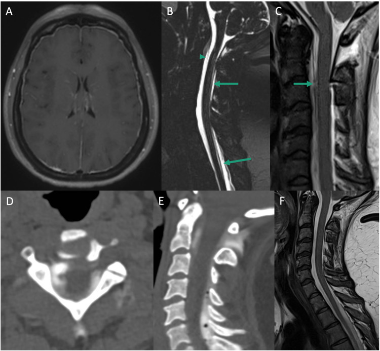
BackgroundSpontaneous intracranial hypotension (SIH) is a severe condition caused by cerebrospinal fluid (CSF) leaks, leading to headaches and neurological impairments. Traditional epidural blood patch treatment is often ineffective, especially in refractory cases without a source leak identified. This study introduces and evaluates the Catheter-Assisted Sealing Technique (CAST), a novel neurointerventional approach for refractory SIH. CAST involves targeted or diffuse delivery of fibrin glue into the epidural space to repair CSF leaks.MethodsA retrospective case series of ten patients with refractory SIH underwent CAST using a fluoroscopy-guided epidural catheter approach. Clinical symptom resolution and follow-up MRI findings were assessed.ResultsNine of 10 patients achieved complete headache resolution and associated symptom relief. One patient experienced significant improvement without complete relief. Follow-up MRI showed resolution of pachymeningeal enhancement in all patients. One patient required repeat procedures for recurrent symptoms. No neurological injury or spinal cord compression occurred, with the only complication being transient femoral nerve palsy due to positioning that resolved within four months.ConclusionCAST is a promising, minimally invasive alternative for refractory SIH. It enables targeted or diffuse fibrin sealant application, providing a seemingly effective leak closure even in cases of occult or multifocal leaks. Early results show high success and safety rates, but larger cohorts and extended follow-up are needed to validate it as a standard treatment.

Keywords: CSF leak; Spine; epidural blood patch; intracranial hypotension.


Conflict of interest statement

The authors declared no potential conflicts of interest with respect to the research, authorship, and/or publication of this article.

Figures

Figure 1.
Figure 1.
Diagram of our institution-specific workup and management of suspected spontaneous CSF leak. SIH: spontaneous intracranial hypotension; MRI: magnetic resonance imaging; w/wo: with, without; CSF: cerebrospinal fluid; SLEC: spinal longitudinal epidural collection; CT: computed tomography; CAST: catheter-assisted sealing of leak technique.
Figure 2.
Figure 2.
Representative intraoperative images during catheter-assisted sealing of leak technique (CAST) procedure on a patient with Type II leak in the C-spine. (A) Fluoroscopic image showing lumbar epidural access with Touhy needle and confirmation with injection of contrasted saline into the epidural space. (B) Fluoroscopic image demonstrating the introduction of a 23 cm 5-French Brite Tip sheath over 0.35 mm glidewire in the lumbar epidural space, being directed cranially. (C) Fluoroscopic image showing the 4-French vertebral navigation in the thoracic epidural space. (D) 0.027 in microcatheter over 0.024 in microwire navigation in the upper cervical epidural space, tip marked by arrow. (E) Negative roadmap view of contrasted dural sealant application, which is seen filling the cervical epidural space. (F) Contrasted dural sealant is seen filling the epidural space, including the C1-2 interspinous space, when compared to image D, microcatheter tip marked with arrow.
Figure 3.
Figure 3.
Imaging from Type I leak. (A) Preoperative axial T1 post-contrast demonstrating diffuse pachymeningeal enhancement and engorgement. (B) Preoperative sagittal cervicothoracic T2 MRI without significant SLEC. (C) Sagittal CT myelogram concerning for CSF leak at dorsal C1/2 interspace (green arrow), posterior cervical SLEC (green arrowheads). (D) Follow-up axial post-contrast T1 MRI with resolution of preoperative meningeal enhancement. (E) Follow-up cervical sagittal T2 MRI reveals a hyperintense collection of vistaseel in the C1/2 interspace, small possible posterior SLEC. (F) Axial post CAST CT reveals contrasted Vistaseel in the dorsolateral epidural space. (G) Sagittal post CAST CT reveals contrasted Vistaseel throughout the cervical epidural space.
Figure 4.
Figure 4.
Imaging from the case of iatrogenic CSF leak after LP. (A) Preoperative axial T2 MRI revealing large, bilateral posterior fossa subdural collections of mixed density with cerebellar compression. (B) Preoperative axial T2 MRI revealing bilateral, mixed-density, convexity subdural collections. (C) Preoperative post-contrast, sagittal T1 MRI demonstrating diffuse meningeal enhancement, posterior fossa crowding, collapsed suprasellar cistern, and reduced mamillopontine distance. (D) Follow-up axial T2 MRI with significantly reduced posterior fossa subdural collections, reduced cerebellar compression. (E) Follow-up axial T2 MRI with resolution of bilateral convexity subdural collections. (F) Postoperative post-contrast, sagittal T1 MRI with resolution of meningeal enhancement, enlarged suprasellar cistern.
Figure 5.
Figure 5.
Imaging from Type 2 (meningeal diverticula) leak. (A) Preoperative axial T1 post-contrast demonstrating diffuse pachymeningeal enhancement and engorgement. (B) Sagittal T2 cervicothoracic MRI where both anterior and posterior SLEC can be visualized (green arrows). (C) Sagittal CT myelogram with anterior and posterior cervicothoracic SLEC (green arrows), but no definitive CSF leak. (D) Follow-up axial T1 post-contrast MRI revealing resolution of meningeal enhancement. (E) Follow-up sagittal T2 cervicothoracic MRI revealing persistent posterior SLEC, resolved anterior cervical SLEC. (F) Sagittal post CAST cervical CT with contrasted Vistaseel identified primarily in the dorsal epidural space. (G) Sagittal post CAST thoracic CT with contrasted Vistaseel in the dorsal epidural space.
Figure 6.
Figure 6.
Imaging from a case with Type I leak. (A) Preoperative MRI axial T1 post contrast showing diffuse meningeal enhancement, reduced ventricular caliber. (B) Preoperative sagittal T2 SPACE cervicothoracic spine demonstrating upper cervical ventral SLEC (arrowhead) and diffuse cervicothoracic dorsal SLEC (arrows). (C) Preoperative sagittal T2 cervical spine in flexed position with enlarged ventral SLEC and area of ventral dural thinning or dehiscence at C2/3 (arrow). (D) Post CAST axial cervical CT showing dural sealant both dorsal and ventral to the thecal sac. (E) Post CAST sagittal cervical CT again showing ventral and dorsal dural sealant in the upper cervical spine. (F) Follow-up sagittal T2 cervical MRI showing resolution of ventral and dorsal SLEC, resolution of CSF signal at C1-2 interspace.

References

    1. D’Antona L, Jaime Merchan MA, Vassiliou A, et al. Clinical presentation, investigation findings, and treatment outcomes of spontaneous intracranial hypotension syndrome: a systematic review and meta-analysis. JAMA Neurol 2021; 78: 329–337. - PMC - PubMed
    1. Schievink WI, Maya MM, Moser FG, et al. Incidence of spontaneous intracranial hypotension in a community: Beverly Hills, California, 2006–2020. Cephalalgia 2022; 42: 312–316. - PubMed
    1. Capizzano AA, Lai L, Kim J, et al. Atypical presentations of intracranial hypotension: comparison with classic spontaneous intracranial hypotension. AJNR Am J Neuroradiol 2016; 37: 1256–1261. - PMC - PubMed
    1. Liaquat MT, Jain S. Spontaneous intracranial hypotension. In: StatPearls [Internet]. Treasure Island (FL): StatPearls Publishing. http://www.ncbi.nlm.nih.gov/books/NBK559066/ (2024, accessed 9 August 2024).
    1. Schievink WI. Misdiagnosis of spontaneous intracranial hypotension. Arch Neurol 2003; 60: 1713–1718. - PubMed

LinkOut - more resources