. 2025 Sep;61(8):991-1004.
doi: 10.1007/s11626-025-01091-6. Epub 2025 Sep 30.

A co-culture system to study the effects of Poly I:C-activated microglia on the differentiation of murine primary neural stem cells

Affiliations

A co-culture system to study the effects of Poly I:C-activated microglia on the differentiation of murine primary neural stem cells

Marie Pierre Manitz et al. In Vitro Cell Dev Biol Anim. 2025 Sep.

Abstract

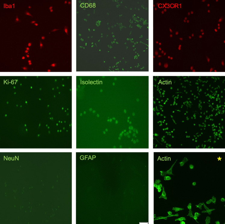
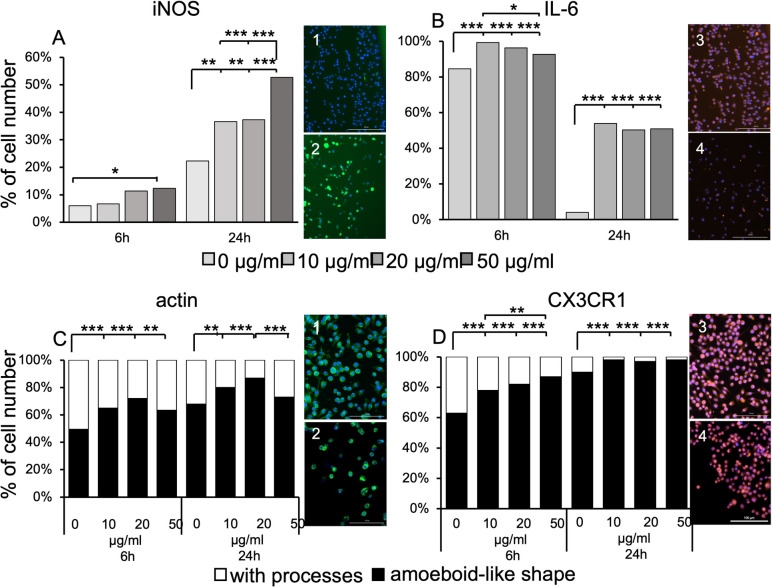
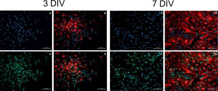
Studies in rodents have shown that systemic inflammation induced by prenatal exposure to the viral mimetic polyinosinic:polycytidylic acid (Poly I:C) triggers maternal immune activation. Cytokines released by the maternal immune system can cross the placenta and enter fetal circulation. In the fetal brain, embryonic microglia may produce additional cytokines and other inflammatory mediators in response to maternally derived cytokines. This resulting cytokine imbalance is suggested to impair neurogenesis and brain development, potentially contributing to the onset of neuropsychiatric disorders in offspring. To investigate microglial involvement in neurogenesis under pathological conditions, we used the spontaneously immortalized microglial cell line (SIM-A9), and confirmed the expression of Iba1 and CD68 via immunocytochemistry. Additionally, SIM-A9 cells expressed CX3CR1, Ki67, and isolectin. Upon Poly I:C stimulation, SIM-A9 cells released the cytokines interleukin-6 (IL-6) and tumor necrosis factor-alpha (TNF-α), as well as nitric oxide (NO), as determined by ELISA and Griess assay, respectively. After confirming SIM-A9 cell activation by Poly I:C, we co-cultured these cells with neural stem/progenitor cells (NSPCs) from embryonic mouse neocortex using a transwell system. We examined how chronically activated microglia influence NSPC differentiation and characterized the resulting cell phenotypes using immunocytochemistry. Our results demonstrate that SIM-A9 cells support NSPC differentiation into neurons as early as three days in culture. However, the number of neurons decreased with prolonged culture. Furthermore, Poly I:C in the NSPC culture media, as well as cytokines secreted by Poly I:C-activated SIM-A9 cells, showed a supportive effect on astrocyte differentiation.

Keywords: Co-culture; Microglia; Neuronal stem cells; Poly I:C.


Conflict of interest statement

Declarations. Ethics approval: All experiments were performed according to the principles regarding the care and use of animals adopted by the German Animal Welfare Law for the prevention of cruelty to animals after approval by the LANUV (Landesamt für Natur, Umwelt und Verbraucherschutz, Nordrhine-Westfalia). This article does not contain any studies involving human participants performed by any of the authors. Conflicts of interest: The authors have no relevant financial or non-financial interests to disclose.

Figures

Figure 1.
Figure 1.
Schematic of experimental design: co-culture of NSPCs and SIM-A9 cells. NSPCs were co-cultured with SIM-A9 cells either for 3 d or 7 d in a transwell-system. SIM-A9 cells were activated with 10 μg/ml or 50 μg/ml Poly I:C to induce production and secretion of inflammatory molecules. NSPCs incubated with Poly I:C (10 μg/ml or 50 μg/ml) and without Poly I:C were used as controls.
Figure 2.
Figure 2.
SIM-A9 cells expressing microglia-specific (Iba1, CD68, and CX3CR1) and non-specific (Isolectin, Actin, and Ki-67) proteins. Immunostaining was performed with SIM-A9 cells (passages 12–14) after 24 h culture. Magnification 200x. There was neither an expression of NeuN nor GFAP. Staining with phalloidin shows the different morphologies of SIM-A9 cells in culture. Asterisk: magnification 400x. Representative fluorescence images of 3 independent experiments.
Figure 3.
Figure 3.
Differential expression and morphological changes in SIM-A9 cells in response to Poly I:C. Cells were exposed to different concentrations (10, 20, and 50 µg/ml) of Poly I:C for 6 and 24 h (h). Nuclei were stained with DAPI (blue). A: Percentage of SIM-A9 cells stained positive for iNOS (green). An increase in iNOS expression was observed in a concentration- and time-dependent manner. a1: SIM-A9 cells exposed to 10 µg/ml Poly I:C for 6 h, showing few or weakly iNOS-positive cells. a2: SIM-A9 cells exposed to 50 µg/ml Poly I:C for 24 h, showing many iNOS-positive cells. Scale bar = 200 µm B: Percentage of SIM-A9 cells positive for IL-6 (red). Prolonged culture led to a decrease of IL-6 expression in SIM-A9 cells. After 6 h, Poly I:C caused an increase in IL-6 expression with the highest IL-6 expression observed at 10 µg/ml Poly I:C). b3: SIM-A9 cells exposed to 10 µg/ml Poly I:C for 6 h, showing IL-6-positive staining. b4: SIM-A9 cells exposed to 20 µg/ml Poly I:C for 24 h, showing few or weakly IL-6-positive cells. Scale bar = 200 µm. C: Percentage of SIM-A9 cells stained for actin with phalloidin (green), showing amoeboid-like morphology (rounded cells without obvious processes; black) or microglial processes (white). Upon Poly I:C stimulation, the number of amoeboid-like SIM-A9 cells increased in a time- and dose-dependent manner, except at the highest concentration. After 24 h of Poly I:C stimulation, the highest number of rounded SIM-A9 cells was observed at 20 µg/ml Poly I:C. Cells exposed to 50 µg/ml Poly I:C showed a higher proportion of rounded cells, but after 24 h, the difference was no longer significant compared to unstimulated cells. c1: Unstimulated SIM-A9 displaying long and short processes cells after 6 h of culture, stained positive for phalloidin. c2: SIM-A9 cells exposed to 20 µg/ml Poly I:C for 24 h, with many amoeboid-like cells stained positive for phalloidin. Scale bar = 100 mm. D: Percentage of SIM-A9 cells stained for CX3CR1 (red), with an amoeboid-like morphology (rounded cells without obvious processes; black) or with microglial processes (white). An increase in rounded cells was observed in a time- and dose-dependent manner. d3: SIM-A9 cells exposed to 10 µg/ml Poly I:C for 6 h, showing some cells with processes. d4: SIM-A9 cells exposed to 50 µg/ml Poly I:C for 24 h, showing many rounded cells. Scale bar = 100 µm. Experiments were performed in four replicates, with approximately 1200 cells counted per staining. * p <.05; ** p <.01; *** p <.001.
Figure 4.
Figure 4.
Effect of Poly I:C stimulation on the secretion of IL-6, TNFα, and NO by SIM-A9 cells analyzed using ELISA and Griess Assay. SIM-A9 cells were cultured in 24 well plate and grown to confluency (~ 90%). After washing cell culture medium without serum containing 10, 20, or 50 µg/ml Poly I:C was added to the cells. After 2, 4, 6, and 24 h 50 µl of the supernatant was removed from each well for ELISA analysis. 4a: In the absence of Poly I:C stimulation (0 µg/ml) IL-6 was not detected in the supernatant at any time point. Increasing Poly I:C concentration (20 and 50 µg/ml) led to increased IL-6 concentrations in a time-dependent manner. 4b: Increasing Poly I:C concentration (10, 20 and 50 µg/ml) led to increased TNF-α production and secretion into supernatant. Maximal TNFα-concentrations were measured after 6 h incubation independently of the Poly I:C concentrations. Prolonged incubation times caused a decrease of TNF-α concentrations. 4c: An increased accumulation of nitrite in cell supernatant was observed in a dose-dependent manner. The concentration of NO production was quantified by being plotted against a standard curve. The experiment was replicated three times. Data represent the mean ± SEM
Figure 5.
Figure 5.
Percentages of neurons, astrocytes, and unlabeled cells after 3 and 7 d of culture. Results were obtained from five independent experiments (5 dams; 41 embryos in total). In each experiment NSPCs were plated in 24-well plates in duplicate for all conditions. Cells cultured for 3 and 7 DIV were stained with anti-GFAP and anti-ßIII-tubulin antibodies to mark astrocytes and neurons, respectively. Cell nuclei were stained with DAPI. A minimum of 500 cells (neurons, astrocytes and unlabeled cells combined) were counted per well. SIM-A9 cells promoted the differentiation of NSPCs into neurons. Poly I:C, at both concentrations and with prolonged culture, promoted the differentiation of NSPCs into astrocytes. Bonferroni post-hoc test was performed to detect differences between the groups of interest. No value = p >.05 (see table).
Figure 6.
Figure 6.
Representative pictures of differentiated astrocytes and neurons derived from NSCs isolated at E13 after three and seven days of co-culture with SIM-A9 cells (N + S). (A) Cell nuclei (in blue) stained with DAPI. (B) Astrocytes (in red) after immunostaining analysis using the anti-GFAP-antibody. (C) Neurons (in green) after immunostaining analysis using the anti-ßIII-tubulin-antibody. (D) merged images (combined red, green, and blue). The highest number of neurons and the lowest number of astrocytes on 3 DIV were observed under condition „N + S “. After 3 DIV, astrocytes displayed an asymmetric morphology with cell processes more or less elongated. After 7 DIV, irrespective of whether they were bipolar or multipolar, they were flattened and extended their surface. This indicates that astrocytes are able to exhibit various cellular shapes in vitro. It seems that despite the presence of SIM-A9 cells, the number of neurons decreased. Scale bar = 100 μm.

References

    1. Aarum J, Sandberg K, Haeberlein SL, Persson MA (2003) Migration and differentiation of neural precursor cells can be directed by microglia. Proc Natl Acad Sci USA 100(26):15983–15988. 10.1073/pnas.2237050100 - PMC - PubMed
    1. Ajmone-Cat MA, Nicolini A, Minghetti L (2003) Prolonged exposure of microglia to lipopolysaccharide modifies the intracellular signaling pathways and selectively promotes prostaglandin E2 synthesis. J Neurochem 87(5):1193–1203. 10.1046/j.1471-4159.2003.02087.x - PubMed
    1. Babon JJ, Varghese LN, Nicola NA (2014) Inhibition of IL-6 family cytokines by SOCS3. Semin Immunol 26(1):13–19. 10.1016/j.smim.2013.12.004 - PMC - PubMed
    1. Baker BJ, Akhtar LN, Benveniste EN (2009) SOCS1 and SOCS3 in the control of CNS immunity. Trends Immunol 30(8):392–400. 10.1016/j.it.2009.07.001 - PMC - PubMed
    1. Bernardino L, Agasse F, Silva B, Ferreira R, Grade S, Malva JO (2008) Tumor necrosis factor-alpha modulates survival, proliferation, and neuronal differentiation in neonatal subventricular zone cell cultures. Stem Cells 26(9):2361–2371. 10.1634/stemcells.2007-0914 - PubMed

LinkOut - more resources