. 2025 Sep 30;15(1):33796.
doi: 10.1038/s41598-025-03890-z.

Sinapic acid accelerates diabetic wound healing by promoting angiogenesis and reducing oxidative stress

Affiliations

Sinapic acid accelerates diabetic wound healing by promoting angiogenesis and reducing oxidative stress

Rupal Dubey et al. Sci Rep. .

Abstract

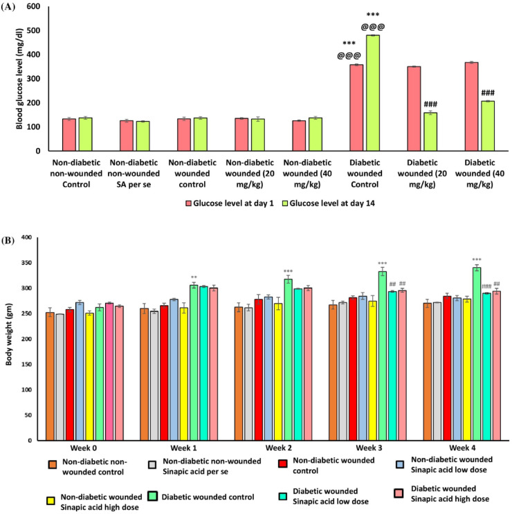
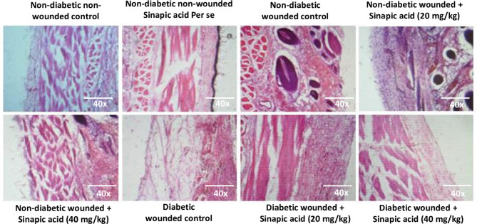
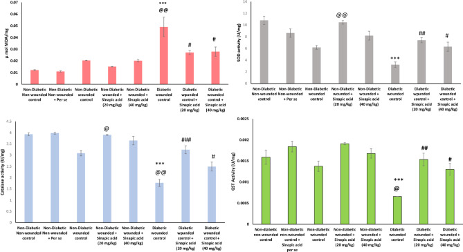
The incidence of delayed wound healing associated with diabetes is increasing globally. Synthetic drugs for wound management often carry adverse effects, underscoring the need for safer and more effective alternatives. Sinapic acid, a phytochemical found in edible plants such as spices, citrus fruits, and berries, has drawn attention for its potential in addressing diabetic wounds. This study investigates the protective effects of sinapic acid at two pharmacological doses in mitigating delayed wound healing and oxidative stress linked to type 2 diabetes. In-vitro, the effects of sinapic acid on cell toxicity, migration, and antioxidant activity were evaluated under high-glucose conditions using L929 murine fibroblast cells and human umbilical vein endothelial cells (HUVEC). In-vivo, diabetic wounds were induced in male Sprague Dawley rats fed with high-fat diet and treated with streptozotocin. Sinapic acid was administered orally at 20 mg/kg and 40 mg/kg, and its efficacy was assessed using an excision wound model. Key markers, including hepatic, and lipid profiles, were analyzed. The findings revealed that sinapic acid significantly improved blood glucose levels and oxidative stress markers in diabetic wound rats. Enhanced angiogenesis, re-epithelialization, wound contraction, cell migration, and SIRT1 levels were observed. This is the first report demonstrating that oral sinapic acid at 20 mg/kg and 40 mg/kg mitigates oxidative stress and promotes wound healing in streptozotocin/high-fat diet (STZ/HFD)-induced diabetes, with lower doses showing greater efficacy.

Keywords: Angiogenesis; Delayed wound healing; Diabetes; Oxidative stress; Sinapic acid.


Conflict of interest statement

Declarations. Competing interests: The authors declare no competing interests.

Figures

Fig. 1
Fig. 1
Schematic representation of the experimental design, diabetic wound induction process, and Sinapic acid treatment.
Fig. 2
Fig. 2
DPPH scavenging activity of (A) Ascorbic acid (B) Sinapic acid. The data is presented as the Mean ± SEM from three independent tests.
Fig. 3
Fig. 3
Cell viability on L929 cells after Sinapic acid treatment. The data is presented as the Mean ± SEM from three independent tests.
Fig. 4
Fig. 4
Effect of sinapic acid on cell migration under hyperglycemic conditions (30 mM glucose). (A) Representative images of wound closure at 0 h and 24 h following treatment with sinapic acid (10 μM and 50 μM). (B) Quantification of % wound closure. Data are presented as mean ± SEM from three independent experiments (n = 3). **p < 0.01, ***p < 0.001 versus high glucose (HG) control.
Fig. 5
Fig. 5
Effect of sinapic acid (10 μM and 50 μM) on angiogenic parameters in HUVECs under hyperglycemic conditions (30 mM glucose). (A) Number of branches, (B) Tube length, and (C) Representative images of tube formation and capillary thickening. Data are expressed as mean ± SEM from three independent experiments (n = 3). *p < 0.05, ***p < 0.001 versus HG control.
Fig. 6
Fig. 6
Effect of Sinapic acid at 100 µM and 150 µM concentrations on antioxidant enzymes under low glucose (LG) and HG conditions using post nuclear supernatant after cell lysis (A) MDA (B) SOD (C) CAT (D) GST. All the values were represented as Mean ± SEM (n = 3), ***p < 0.001, versus Control LG and ###p < 0.001, versus HG control.
Fig. 7
Fig. 7
Effect of Sinapic acid on 20 mg/kg and 40 mg/kg on (A) Blood glucose level (B) Body weight. The data is represented as Mean ± SEM. ***represents p < 0.001, when in comparison with the non-diabetic non-wounded control group; @@@represents p < 0.001, when in comparison with the non-diabetic wounded control group; ###represents p < 0.001, when in comparison to the diabetic wounded control group.
Fig. 8
Fig. 8
Effect of Sinapic acid on wound healing parameters (A) % Wound closure (B) Photographic representation of wound contraction on day 0, 7, and 14. The data is represented as Mean ± SEM. @@@represents p < 0.001, @@represents p < 0.01, and @represents p < 0.05, when in comparison to the non-diabetic wounded control group; ###represents p < 0.001; ##represents p < 0.01, and #represents p < 0.05, when in comparison to the diabetic wounded control group.
Fig. 9
Fig. 9
Histopathological evaluation of the diabetic wound healing process before and after treatment in different experimental groups, captured at 40 × magnification.
Fig. 10
Fig. 10
Effect of Sinapic acid at 20 mg/kg and 40 mg/kg on lipid markers (A) Cholesterol (B) Triglycerides (C) VLDL (D) LDL (E) HDL. The data is represented as Mean ± SEM. ***represents p < 0.001, *represents p < 0.05; when in comparison with the non-diabetic non-wounded control group; @@@represents p < 0.001; @@represents p < 0.01; @represents p < 0.5, when in comparison to the non-diabetic wounded control group; ###represents p < 0.001; ##represents p < 0.01; #represents p < 0.5, when in comparison to the diabetic wounded control group.
Fig. 11
Fig. 11
Effect of Sinapic acid at 20 mg/kg and 40 mg/kg on hepatotoxicity markers (A) SGPT (B) SGOT (C) Bilirubin (D) Alkaline phosphatase. The data is represented as Mean ± SEM. ***represents p < 0.001, **represents p < 0.01; *represents p < 0.5, when in comparison with the non-diabetic non-wounded control group; @@@represents p < 0.001, @represents p < 0.5, when in comparison with the non-diabetic wounded control group; ###represents p < 0.01; ##represents p < 0.01; #represents p < 0.5, when in comparison to the diabetic wounded control group.
Fig. 12
Fig. 12
Effect of Sinapic acid at 20 mg/kg and 40 mg/kg in skin homogenate (A) LPO (B) SOD (C) CAT (D) GST (E) GSH. The data is represented as Mean ± SEM. ***represents p < 0.001; **p < 0.01, *represents p < 0.5, when in comparison with the non-diabetic non-wounded control group; @@@represents p < 0.001; @@p < 0.01, @represents p < 0.5, when in comparison with the non-diabetic wounded control group; ###represents p < 0.001; ##represents p < 0.01; #represents p < 0.5, when in comparison to the diabetic wounded control group.
Fig. 13
Fig. 13
Effect of Sinapic acid at 20 mg/kg and 40 mg/kg on concentration of SIRT1. The data is represented as Mean ± SEM. ***represents p < 0.001, when in comparison with the non-diabetic non-wounded control group; @@@represents p < 0.001, @@represents p < 0.01, when in comparison with the non-diabetic wounded control group; ###represents p < 0.001, when in comparison to the diabetic wounded group.

References

    1. IDF Diabetes Atlas, 11th edn. Brussels, Belgium: International Diabetes Federation (2025).
    1. Raghav, A. et al. Financial burden of diabetic foot ulcers to world: A progressive topic to discuss always. Ther. Adv. Endocrinol. Metab.9, 29–31 (2018). - PMC - PubMed
    1. Yang, D.-R., Wang, M.-Y., Zhang, C.-L. & Wang, Y. Endothelial dysfunction in vascular complications of diabetes: A comprehensive review of mechanisms and implications. Front. Endocrinol. (Lausanne)15, 1359255 (2024). - PMC - PubMed
    1. Huang, K. et al. Angiogenesis during diabetic wound repair: From mechanism to therapy opportunity. Burns Trauma13, tkae052 (2025). - PMC - PubMed
    1. Li, Y. et al. Diabetic vascular diseases: Molecular mechanisms and therapeutic strategies. Signal Transduct. Target. Ther.8, 152 (2023). - PMC - PubMed

MeSH terms

LinkOut - more resources