Case Reports
. 2025 Sep 15:63:103212.
doi: 10.1016/j.eucr.2025.103212. eCollection 2025 Nov.

Paratesticular myxofibrosarcoma in an adult male: A rare case in an unusual anatomical site

Affiliations
Case Reports

Paratesticular myxofibrosarcoma in an adult male: A rare case in an unusual anatomical site

Yacob Sheiferawe Seman et al. Urol Case Rep. .

Abstract

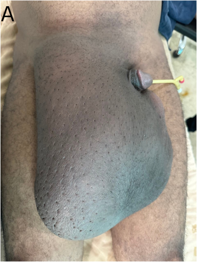
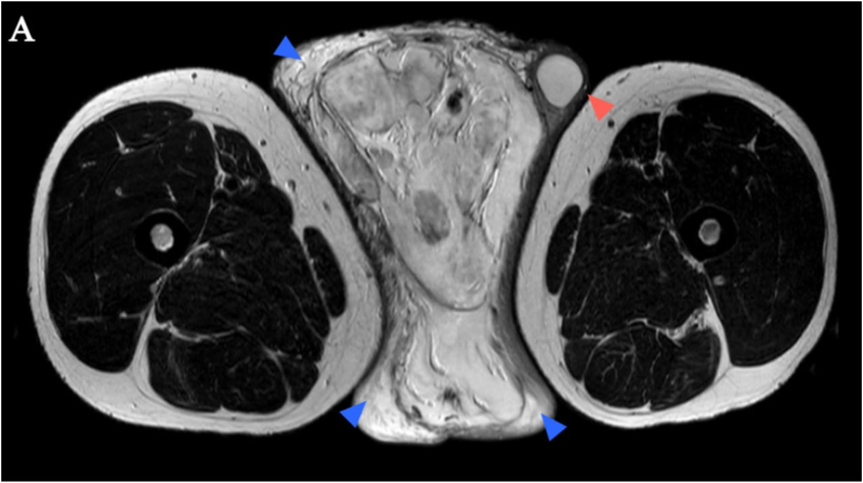
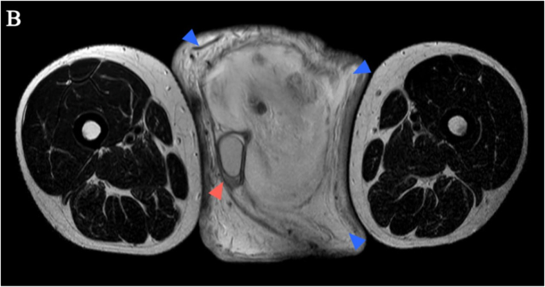
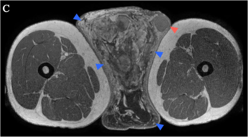
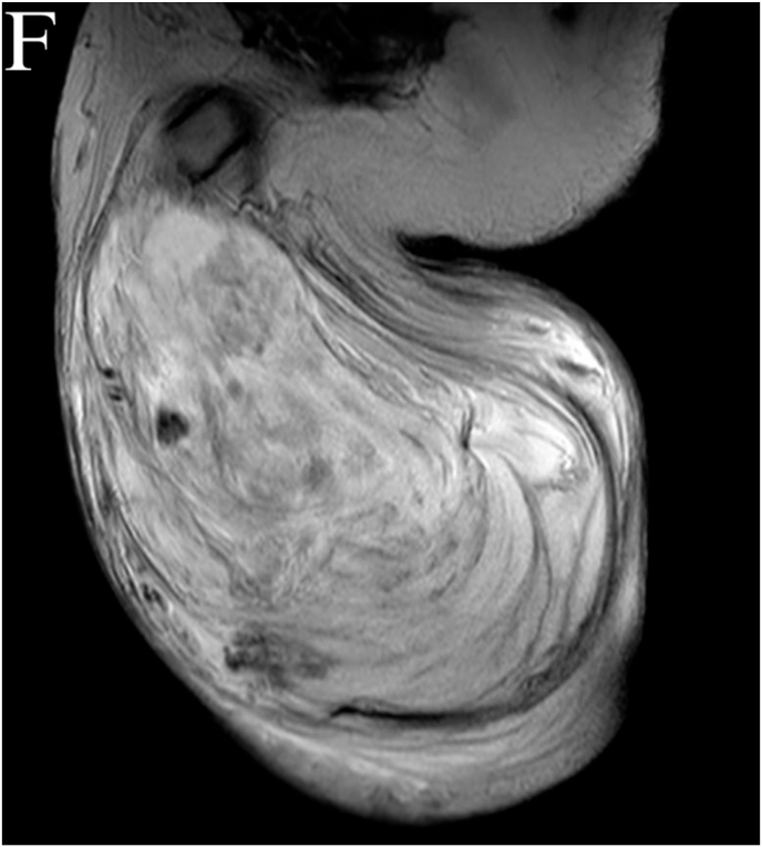
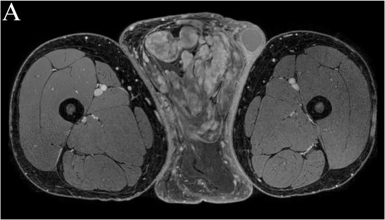
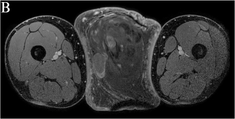
Myxofibrosarcoma is a rare malignant soft tissue tumor, most commonly affecting the extremities of elderly individuals. Paratesticular involvement is extremely uncommon and often presents a diagnostic challenge. We present the case of a male patient who presented with a painless, progressively enlarging right-sided scrotal swelling of 4 years' duration. Clinical examination revealed a large scrotal mass with a non-palpable right testicle. Imaging and histopathological evaluation confirmed the diagnosis of paratesticular myxofibrosarcoma. We will mainly discuss the clinical presentation, imaging findings, and pathological features of this rare tumor.

Keywords: Magnetic resonance imaging; Myxofibrosarcoma; Paratesticular tumor; Scrotal mass.


Conflict of interest statement

The authors declare that they have no known competing financial interests or personal relationships that could have appeared to influence the work reported in this paper.

Figures

Fig. 1
Fig. 1
Pre-operative pictures of the scrotal lesion in supine (A) and standing (B) positions.
Fig. 1
Fig. 1
Pre-operative pictures of the scrotal lesion in supine (A) and standing (B) positions.
Fig. 2
Fig. 2
Axial Pelvic MRI in T2 (A, B), T1 (C, D), T2 coronal (E), and T2 sagittal (F) sequences: They show a large (24cm × 20cm x 14cm- CCxAPxTR) heterogeneous infiltrative right hemi-scrotal mass (blue arrowheads in A-D). The lesion shows predominantly hyperintense signal on T2 and hypointense signal on T1. The right and left testes (red arrowheads in A-D) are splayed peripherally but have a sharp border against the mass. The epididymis is not separately seen. The coronal (E) and sagittal (F) T2 images show the scrotal skin thickening to a better advantage.
Fig. 2
Fig. 2
Axial Pelvic MRI in T2 (A, B), T1 (C, D), T2 coronal (E), and T2 sagittal (F) sequences: They show a large (24cm × 20cm x 14cm- CCxAPxTR) heterogeneous infiltrative right hemi-scrotal mass (blue arrowheads in A-D). The lesion shows predominantly hyperintense signal on T2 and hypointense signal on T1. The right and left testes (red arrowheads in A-D) are splayed peripherally but have a sharp border against the mass. The epididymis is not separately seen. The coronal (E) and sagittal (F) T2 images show the scrotal skin thickening to a better advantage.
Fig. 3
Fig. 3
Axial T1 C+ pelvic MRI: The lesion shows significant enhancement especially in its more proximal part (A) compared to its distal part (B).
Fig. 3
Fig. 3
Axial T1 C+ pelvic MRI: The lesion shows significant enhancement especially in its more proximal part (A) compared to its distal part (B).
Fig. 4
Fig. 4
Post-operative pictures of the resected mass lesion are seen (A) and (B). The mass had areas of necrosis and hemorrhage with multinodular growth pattern on gross examination. A Foley catheter is placed adjacently as a measurement scale.
Fig. 4
Fig. 4
Post-operative pictures of the resected mass lesion are seen (A) and (B). The mass had areas of necrosis and hemorrhage with multinodular growth pattern on gross examination. A Foley catheter is placed adjacently as a measurement scale.
Fig. 5
Fig. 5
Hematoxylin and Eosin Stains in x10 (A, B) and x40 (C) powers: Hypocellular and hypercellular areas composed of spindle to stellate cells admixed with elongated, curvilinear blood vessels in a myxoid background consistent with MFS.
Fig. 5
Fig. 5
Hematoxylin and Eosin Stains in x10 (A, B) and x40 (C) powers: Hypocellular and hypercellular areas composed of spindle to stellate cells admixed with elongated, curvilinear blood vessels in a myxoid background consistent with MFS.
Fig. 5
Fig. 5
Hematoxylin and Eosin Stains in x10 (A, B) and x40 (C) powers: Hypocellular and hypercellular areas composed of spindle to stellate cells admixed with elongated, curvilinear blood vessels in a myxoid background consistent with MFS.

References

    1. Roland C.L., Wang W.L., Lazar A.J., Torres K.E. Myxofibrosarcoma. Surg Oncol Clin N Am. 2016 Oct;25(4):775–788. doi: 10.1016/j.soc.2016.05.008. Epub 2016 Jul 30. PMID: 27591498. - DOI - PubMed
    1. Elias A., Ryan L., Sulkes A., Collins J., Aisner J., Antman K.H. Response to mesna, doxorubicin, ifosfamide, and dacarbazine in 108 patients with metastatic or unresectable sarcoma and no prior chemotherapy. J Clin Oncol. 1989 Sep;7(9):1208–1216. doi: 10.1200/JCO.1989.7.9.1208. PMID: 2504890. - DOI - PubMed
    1. Merck C., Angervall L., Kindblom L.G., Odén A. Myxofibrosarcoma. A malignant soft tissue tumor of fibroblastic-histiocytic origin. A clinicopathologic and prognostic study of 110 cases using multivariate analysis. Acta Pathol Microbiol Immunol Scand Suppl. 1983;282:1–40. PMID: 6444190. - PubMed
    1. Russo P., Brady M.S., Conlon K., et al. Adult urological sarcoma. J Urol. 1992 Apr;147(4):1032–1036. doi: 10.1016/s0022-5347(17)37456-6. discussion 1036-7 PMID: 1552580. - DOI - PubMed
    1. Respati E., Prapiska F.F., Sihombing B., Warli S.M., Hartati L., Adelaidey A.N. First reported case of adult paratesticular myxofibrosarcoma in Indonesia: case report and literature review. Int J Surg Case Rep. 2022 Apr;93 doi: 10.1016/j.ijscr.2022.106849. Epub 2022 Feb 25. PMID: 35286976; PMCID: PMC8921302. rad. - DOI - PMC - PubMed

Publication types

LinkOut - more resources