. 2025 Oct;51(5):e70041.
doi: 10.1111/nan.70041.

Use of DNA Methylation Profiling as a Molecular Classification Tool for Paediatric Central Nervous System Tumours: A Middle-Income Country Population-Based Study

Affiliations

Use of DNA Methylation Profiling as a Molecular Classification Tool for Paediatric Central Nervous System Tumours: A Middle-Income Country Population-Based Study

Mayara F Euzébio et al. Neuropathol Appl Neurobiol. 2025 Oct.

Abstract

Paediatric central nervous system (CNS) tumours are the second most common childhood malignancy and the leading cause of cancer-related mortality in this age group. Histopathological diagnosis can be challenging, particularly for rare or ambiguous tumours, and may result in misclassification. To evaluate the utility of DNA methylation profiling in a middle-income country, we performed the Infinium MethylationEPIC BeadChip (Illumina) on tumours from 182 paediatric patients treated at a reference centre for paediatric oncology in Campinas, state of São Paulo, Brazil. Data were analysed using the DKFZ/Heidelberg CNS tumour classifier (v12.8). After excluding control tissue, 163 samples (89.6%) were suitable for classification; 135 (74.2%) achieved a calibrated score ≥ 0.9 and were assigned to a methylation class family. Methylation profiling resulted in a tumour subtype for 88 cases (65.7%) and changed the diagnosis in 28 cases (20.9%), identifying several rare tumour subtypes that were identified solely through methylation analysis, confirming the value of this method in improving diagnostic accuracy. This study highlights the utility of DNA methylation profiling for paediatric CNS tumours in a resource-limited setting and provides a cohort from an underrepresented middle-income population to international molecular databases.

Keywords: DNA methylation profiling; middle‐income countries; molecular classification; molecular tumour subtypes; paediatric CNS tumours.


Conflict of interest statement

The authors declare no conflicts of interest.

Figures

FIGURE 1
FIGURE 1
A) Overview of the molecular classification of the 182‐paediatric CNS tumours cohort. Excluding cases classified as control tissue (n = 19), 135 out of 163 (82.8%) samples were assigned to a methylation family with a high calibrated score of ≥ 0.9. Regarding methylation subclasses, 134 cases (99.3%) achieved a prediction accuracy with a calibrated score of 0.5 or higher. Of these, 106 patients had an initial diagnosis consistent with the molecular classification, while 28 showed discrepancies. In 87 cases, the molecular classification refined the diagnosis by adding a subclass. B) Estimated cell purity (percentage) in relation to the calibrated score. **** p < 0.0001.
FIGURE 2
FIGURE 2
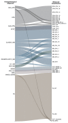
Alluvial plot showing 88 tumours refined by molecular classification (left—initial diagnosis; right—methylation‐based molecular classification). ANA_EPN, anaplastic ependymoma; EPN, ependymoma; SUB_EPN, subependymoma; AT/RT, atypical teratoid rhabdoid tumour; CLASSIC_MB, classic medulloblastoma; DESMOPLASTIC_MB, desmoplastic medulloblastoma; LG_MB, large cells medulloblastoma; NOD_MB, nodular medulloblastoma; GERM, germinoma; ANG_GLI, angiocentric glioma; MEN, meningioma; MIXED_GERM, mixed germ cells tumour; PA, pilocytic astrocytoma; EPN_PFA_1A, Posterior fossa group A (PFA) ependymoma, subclass 1a; EPN_PFA_1B, posterior fossa group A (PFA) ependymoma, subclass 1b; EPN_PFA_1C, posterior fossa group A (PFA) ependymoma, subclass 1c; EPN_PFA_1D, posterior fossa group A (PFA) ependymoma, subclass 1d; EPN_PFA_1F, posterior fossa group A (PFA) ependymoma, subclass 1f; EPN_PFA_2A, posterior fossa group A (PFA) ependymoma, subclass 2a; EPN_PFA_2B, posterior fossa group A (PFA) ependymoma, subclass 2b; EPN_PFB_4, posterior fossa group B ependymoma, subclass 4; EPN_SPINE_SE_A, spinal subependymoma, subtype A; EPN_ST_ZFTA_RELA_A, supratentorial ependymoma, ZFTA fusion‐positive, subtype ZFTA‐RELA fused, subclass A; EPN_YAP, supratentorial ependymoma, YAP1‐fused; ATRT_MYC, atypical teratoid rhabdoid tumour, MYC activated; ATRT_SHH, atypical teratoid rhabdoid tumour, SHH activated; MB_G34_I, Medulloblastoma Group 3, subclass I; MB_G34_II, Medulloblastoma Group 3, subclass II; MB_G34_III, Medulloblastoma Group 3, subclass III; MB_G34_IV, Medulloblastoma Group 3, subclass IV; MB_G34_V, medulloblastoma Group 4, subclass V; MB_G34_VII, medulloblastoma Group 4, subclass VII; MB_G34_VIII, medulloblastoma Group 4, subclass VIII; MB_SHH_1, medulloblastoma, SHH‐activated, subtype 1; MB_SHH_2, medulloblastoma, SHH‐activated, subtype 2; MB_SHH_3, medulloblastoma, SHH‐activated, subtype 3; MB_SHH_4, medulloblastoma, SHH‐activated, subtype 4; MB_WNT, medulloblastoma, WNT activated; GCT_GERM_A, germinoma, subtype A; GCT_GERM_KIT, germinoma, subtype KIT mutant (novel); AG_MYB, angiocentric glioma, MYB/MYBL1‐altered; MNG_BEN_1, meningioma, subclass benign 1; MNG_BEN_3, meningioma, subclass benign 3; PA_INF, infratentorial pilocytic astrocytoma; PA_MID, supratentorial midline pilocytic astrocytoma; GCT_YOLKSAC, yolk sac tumour; PB_GRP2, pineoblastoma, miRNA pathway altered, group 2.
FIGURE 3
FIGURE 3
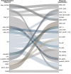
Alluvial plot showing 28 cases that were discrepant with the initial diagnosis and molecular classification (left—initial diagnosis; right—methylation‐based molecular classification). ANA_ASTRO, anaplastic astrocytoma; PA, pilocytic astrocytoma; CNS_ET, CNS embryonal tumour; ANA_EPN, anaplastic ependymoma; SUB_EPN, subependymoma; ANA_GG, anaplastic ganglioglioma; GG, ganglioglioma; NEUROC, central neurocytoma; CPC, choroid plexus carcinoma; CLASSIC_MB, classic medulloblastoma; CRANIOF, craniopharyngioma; DNET, dysembryoplastic neuroepithelial tumour; MULT_GLIO, multiform glioblastoma; PINEO, pineocytoma; DMG_K27, diffuse midline glioma, H3K27‐altered, subtype H3K27‐mutant or EZHIP expressing; ATRT_SHH, atypical teratoid rhabdoid tumour, SHH activated; ATRT_TYR, atypical teratoid rhabdoid tumour, Tyrosinase activated; CNS_NB_FOXR2, CNS neuroblastoma, FOXR2‐altered; DGONC, Diffuse glioneuronal tumour with oligodendroglioma‐like features and nuclear clusters; DMG_EGFR, diffuse midline glioma, H3K27‐altered, subtype EGFR‐altered; GNT_A, Diffuse glioneuronal tumour, subtype A; LGG_MYB_B, diffuse astrocytoma, MYB or MYBL1‐altered, subtype B [infratentorial] (novel); NET_PLAGL1_FUS, neuroepithelial tumour, PLAGL1‐fused; PA_CORT, supratentorial pilocytic astrocytoma; PA_INF, infratentorial pilocytic astrocytoma; PA_MID, supratentorial midline pilocytic astrocytoma; PB_FOXR2, pineoblastoma, MYC/FOXR2‐activated; PB_GRP1A, pineoblastoma, miRNA pathway altered, group 1A; pedHGG_MYCN, diffuse paediatric‐type high‐grade glioma, MYCN subtype; PXA, pleomorphic xanthoastrocytoma; RGNT, rosette‐forming glioneuronal tumour.
FIGURE 4
FIGURE 4
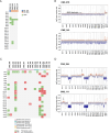
A) Oncoplot showing copy number alterations in all pilocytic astrocytomas with nonflat copy number profiling. PA_INF, infratentorial pilocytic astrocytoma; PA_MID, supratentorial midline pilocytic astrocytoma. B) Representative copy number of tumours with and without BRAF duplication, respectively. Arrow: BRAF duplication. C) Oncoplot showing the CNAs for 16 ependymomas, grouped according to the molecular subclasses. D) Copy number plots of tumours classified as ZFTA::RELA fusion‐positive ependymomas. Arrows indicate chromosomal changes usually found in the supratentorial group.
FIGURE 5
FIGURE 5
A) Copy number plot of a YAP1‐fused supratentorial ependymoma showing copy number aberration around the YAP1 locus (arrow), as expected. B) Copy number plot of CNS_020, reclassified as pineoblastoma, miRNA pathway altered group 2 subtype, showing several chromosome losses. C) Copy number plots of initially diagnosed CNS embryonal tumours. CNS_014 and CNS_043 had chromosome 16 loss and chromosomes 7 and 12 gain, common features of miR pathway altered group 1A subclass; CNS_025 displayed chromosome 8q duplication, typical of MYC/FOXR2‐activated subclass.
FIGURE 6
FIGURE 6
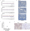
A) Copy number plot showing three tumours classified as AT/RT. CNS_192 and CNS_199 showed SMARCB1 and/or chromosome 21 deletion. B) The same three cases presented an absence of INI1 by immunohistochemistry, supporting the new tumour subtype. Tumour vessels were positive for INI1, considered an internal control for the reaction. C) H3K27M status assessed by ddPCR in the tumour tissue. The scatter plot represents the mutant allele frequency (MAF). Two cases showed high MAF: 25% and 73%, and CNS_065 was not mutated (0%, similar to NC). MAF (%) was calculated as mutant copies per μL/total copies per μL × 100, with the total copies representing the sum of mutant and wild‐type copies. NTC, nontarget control; NC, negative control. D) CNS_065 Immunohistochemical staining for H3K27me3 of case CNS_065 shows reduced expression compared to the control sample.

References

    1. Damodharan S. and Puccetti D., “Pediatric Central Nervous System Tumor Overview and Emerging Treatment Considerations,” Brain Sciences 13, no. 7 (2023): 1106. - PMC - PubMed
    1. Lam C. G., Howard S. C., Bouffet E., and Pritchard‐Jones K., “Science and Health for All Children With Cancer,” Science 363, no. 6432 (2019): 1182–1186. - PubMed
    1. Bertuccio P., Alicandro G., Malvezzi M., et al., “Childhood Cancer Mortality Trends in Europe, 1990–2017, With Focus on Geographic Differences,” Cancer Epidemiology 67 (2020): 101768. - PubMed
    1. Bailey S., Davidson A., Parkes J., et al., “How Can Genomic Innovations in Paediatric Brain Tumours Transform Outcomes in Low‐ and Middle‐Income Countries?,” JCO Global Oncology 8 (2022): e2200156, 10.1200/GO.22.00156. - DOI - PMC - PubMed
    1. da Costa M. D. S., Torres Soares C., Dastoli P. A., et al., “Survival Analysis and Prognostic Factors in Posterior Fossa Ependymomas in Children and Adolescents,” Journal of Neurosurgery. Pediatrics 5, no. 3 (2023): 1–9, 10.3171/2023.5.PEDS22350. - DOI - PubMed

LinkOut - more resources