. 2025 Jun 16;33(3):201007.
doi: 10.1016/j.omton.2025.201007. eCollection 2025 Sep 18.

CircSPINT2 confers sensitivity to osimertinib via hsa-miR-1296-3p/RBP1 axis and inhibits NSCLC progression

Affiliations

CircSPINT2 confers sensitivity to osimertinib via hsa-miR-1296-3p/RBP1 axis and inhibits NSCLC progression

Nalini Devi Verusingam et al. Mol Ther Oncol. .

Abstract

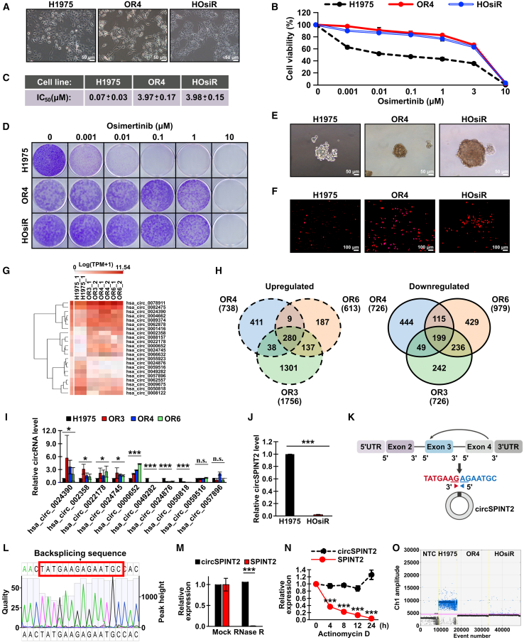
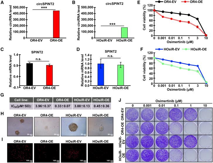
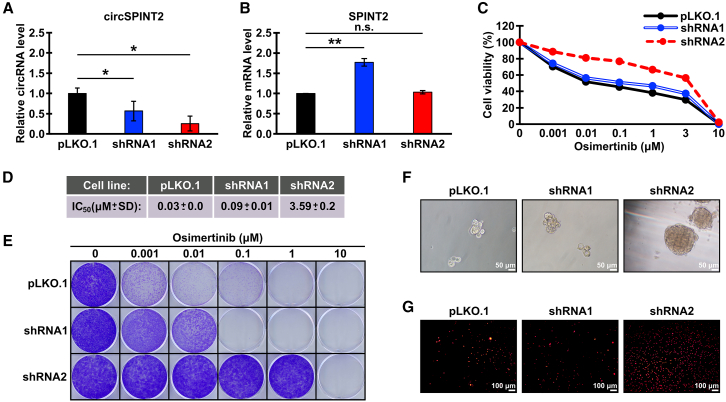
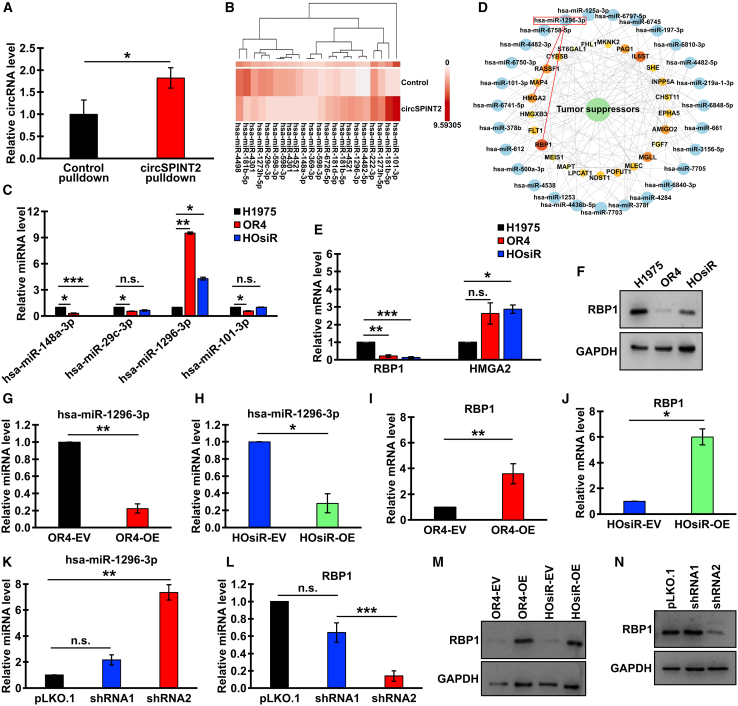
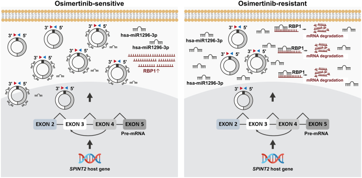
Lung adenocarcinoma (LUAD) is the most common type of lung cancer. Prolonged treatment of LUAD with 1st/2nd generation epidermal growth factor receptor tyrosine kinase inhibitors (EGFR-TKIs) promotes the emergence of secondary EGFR T790M mutation conferring resistance to these drugs. Patients who acquire EGFR T790M mutation respond to the 3rd generation EGFR-TKI osimertinib but develop resistance within 12 months. Circular RNAs (circRNAs) are notably associated with cancerogenesis, making them promising biomarkers or therapeutic targets. In this study, we aimed to identify circRNAs that regulate osimertinib resistance and elucidate the functions and mechanisms to reveal potential therapeutic targets and biomarkers for osimertinib-resistant LUAD. The analysis of circRNA transcriptome sequencing identified circSPINT2 to be downregulated in osimertinib-resistant cell lines. The loss-/gain-of-function assays revealed that circSPINT2 sensitizes cells to osimertinib treatment by inducing apoptosis. Functionally, circSPINT2 enhanced the expression of RBP1 by sponging hsa-miR-1296-3p. Osimertinib-resistant xenograft tumor model was established by long-term osimertinib treatment, and the molecular and histologic analysis of subcutaneous xenograft tumors corroborated our in vitro findings. Conclusively, our study demonstrates that circSPINT2 possesses tumor-suppressive functions; the restoration of circSPINT2 expression level confers sensitivity to osimertinib treatment via miR-1296-3p/RBP1 axis. The secreted circSPINT2 may serve as a monitoring biomarker for osimertinib resistance status in LUAD.

Keywords: EGFR-TKI; L858R; NSCLC; T790M; biomarker; circular RNA; lung cancer; non-coding RNA; osimertinib; osimertinib-resistant.


Conflict of interest statement

The authors declare that they have no conflicts of interest.

Figures

None
Graphical abstract
Figure 1
Figure 1
circSPINT2 is downregulated in osimertinib-resistant cell lines (A) Bright-field images (20× magnification) showing the morphology of osimertinib-resistant cells (OR4 and HOsiR) in comparison with parental H1975 cells. (B) alamarBlue assay showing the viability of H1975, OR4, and HOsiR cells at different concentrations of osimertinib. Data are presented as the percentages of survived cells after 48-h treatment with the indicated concentrations of osimertinib relative to untreated control. (C) The measurements of IC50 of osimertinib against H1975, OR4, and HOsiR cells. (D) Clonogenic assay showing the capacity of H1975, OR4, and HOsiR cells to form colonies at the indicated concentrations of osimertinib. (E) Bright-field images showing tumorspheres formed by H1975, OR4, and HOsiR cell lines. (F) Fluorescent images of transwell migration assay demonstrating the migration capacity of H1975, OR4, and HOsiR cells. (G) Hierarchical clustering heatmap showing the expression of circRNAs in parental (H1975) and OR cell lines. (H) Venn diagrams showing the numbers of unique and common differentially expressed circRNAs in three OR cell lines. (I) RT-qPCR validation of the expression of selected differentially expressed circRNAs in parental and three OR cell lines. (J) RT-qPCR showing the expression of circSPINT2 in HOsiR cells. (K) Schematic of circSPINT2 formation via backsplicing and the splice junction sequence. (L) Sanger sequencing result showing the backsplicing junction of circSPINT2. (M) RT-qPCR amplification of SPINT2 mRNA and circSPINT2 after the treatment of H1975 total RNA with RNase R. (N) RT-qPCR analysis of the stability of SPINT2 and circSPINT2 transcripts in a time course of actinomycin D treatment of H1975 cells. (O) RT-ddPCR analysis showing the level of circSPINT2 secreted into the medium by the indicated cell lines. NTC, no template control. Quantitative data are presented as the means ± SEM, n = 3, ∗p < 0.05, ∗∗p < 0.01 and ∗∗∗p < 0.001 (paired t test).
Figure 2
Figure 2
Overexpression of circSPINT2 enhances osimertinib sensitivity and reduces NSCLC cell aggressiveness (A–D) RT-qPCR showing the expression of circSPINT2 (A and B) and linear SPINT2 mRNA (C and D) in OR4 (A and C) or HOsiR (B and D) cells transfected with empty vector (EV) or circSPINT2 overexpression construct (OE). The mean values relative to EV are shown, n = 3, SEM error bars, ∗∗∗p < 0.001, n.s., not significant (paired t test). (E and F) alamarBlue assay showing the viability of OR4-EV and OR-OE (E) or HOsiR-EV and HOsiR-OE (F) cells at different concentrations of osimertinib. (G) The measurements of IC50 of osimertinib against the indicated cell lines. (H) Bright-field images of tumorspheres formed by the indicated cell lines. (I) Fluorescent images of transwell migration assays of the indicated cell lines. (J) Colony formation assay of OR4 and HOsiR clones at the indicated concentrations of osimertinib. Quantitative data are presented as the means ± SEM error bars, n = 3, ∗p < 0.05, ∗∗p < 0.01, ∗∗∗p < 0.001 using paired t test.
Figure 3
Figure 3
The knockdown of circSPINT2 in H1975 cells promotes osimertinib resistance and enhances NSCLC cell tumorigenicity in vitro (A and B) RT-qPCR showing the expression of circSPINT2 (A) and linear SPINT2 mRNA (B) in H1975 cells stably transfected with circSPINT2-targeting shRNAs (1 and 2). Mean fold changes relative to vector control (pLKO.1) are shown, n = 3, SEM error bars, ∗p < 0.05, ∗∗p < 0.01, n.s., not significant (paired t test). (C) alamarBlue assay showing cell viability in H1975 clones with circSPINT2 knockdown (shRNA1, shRNA2) and control (pLKO.1) treated with increasing concentrations of osimertinib. (D) IC50 values of osimertinib in H1975 cell clones. (E) Colony formation assay of the indicated H1975 cell clones at specified concentrations of osimertinib. (F) Bright-field images of tumorspheres formed by the indicated H1975 cell clones. (G) Fluorescent images of transwell migration assay demonstrating the migration capacity of the indicated H1975 cell clones.
Figure 4
Figure 4
CircSPINT2 promotes apoptotic stress upon osimertinib treatment (A–C) Representative flow cytometry profiles showing the 7-AAD-labeled apoptotic cells after osimertinib treatment in the parental H1975, OR4 and HOsiR cells (A), OR4 and HOsiR cells overexpressing circSPINT2 (OE) as compared to empty vector (EV) control (B), and H1975 cells with the knockdown of circSPINT2 (shRNA1 and shRNA2) as compared to vector control (pLKO.1) (C). (D–F) Quantification of apoptotic cells from flow cytometry data shown in (A–C), respectively. Mean values are shown ±SEM, n = 3, ∗p < 0.05, ∗∗∗p < 0.001 (paired t test). (G) Micro-western array blots showing the expression of the indicated apoptotic markers in the indicated LUAD clones. (H) Quantification of micro-western array (G) visualized as a heatmap.
Figure 5
Figure 5
CircSPINT2 upregulates RBP1 expression by sponging miR-1296-3p (A) RT-qPCR analysis showing circSPINT2 enrichment in pull-down fractions compared to control. (B) Hierarchical clustering heatmap of miRNAs differentially enriched in circSPINT2 pull-down samples. (C) RT-qPCR analysis of selected miRNAs pulled down via circSPINT2 in H1975, OR4, and HOsiR cells. (D) STRING network of predicted interactions between pulled-down miRNAs and target tumor suppressor genes; interactions involving miR-1296-3p are highlighted. (E) RT-qPCR analysis of RBP1 expression in H1975, OR4, and HOsiR cells. (F) Western blots showing the expression of RBP1 protein in H1975, OR4, and HOsiR cells. (G and H) RT-qPCR analysis of hsa-miR-1296 expression in OR cell lines overexpressing circSPINT2 (OE) compared to empty vector controls (EV). (I and J) RT-qPCR analysis of RBP1 mRNA expression in OR cell lines overexpressing circSPINT2 (OE) compared to EV controls. (K and L) RT-qPCR analysis of hsa-miR-1296-3p (K) and RBP1 mRNA (L) expression in H1975 cells with circSPINT2 knockdown (shRNA1 and shRNA2) compared to vector control (pLKO.1). (M and N) Western blots showing RBP1 protein expression in OR cell lines overexpressing circSPINT2 (OE) and EV controls (M) and H1975 cells with circSPINT2 knockdown (shRNA1 and shRNA2) and control (pLKO.1) (N). GAPDH, loading control. Quantitative data for RT-qPCR are presented as the relative mean values ±SEM error bars, n = 3, ∗p < 0.05, ∗∗p < 0.01 and ∗∗∗p < 0.001 (paired t test).
Figure 6
Figure 6
In vivo and ex vivo osimertinib-resistant models demonstrate the downstream signaling networks underlying circSPINT2-mediated drug sensitivity (A) Schematic representation of the experimental setup. H1975 cells were subcutaneously injected into nude mice to form xenograft prior to resistance induction with osimertinib (5 mg/kg). (B) Tumor growth curves from xenografts in the indicated experimental groups. Arrows indicate the start of the treatment and harvesting time of vehicle and osimertinib-treated tumors after reaching ca. 1,000 mm3 in size. (C–E) RT-qPCR analysis of the expression of circSPINT2 (C), RBP1 (D), and miR-1296-3p (E) in xenograft tumors of vehicle and treatment groups. (F) FISH analysis showing the levels and localization of circSPINT2 within the cytoplasm in subcutaneous tumors. Nuclei stained with DAPI. (G) Immunoblot analysis of the expression of RBP1 in the vehicle and osimertinib-treated xenografts. Triplicate samples are shown for each group (L1-L6), GAPDH, loading control. (H) IHC staining of RBP1 in the vehicle and osimertinib-treated xenografts. (I) alamarBlue cell viability assay showing the response to osimertinib of primary cell lines established from subcutaneous tumors derived from vehicle and osimertinib-treated groups. (J) Flow cytometry profiles showing the percentages of 7-AAD-labeled apoptotic cells among the primary cell lines established from subcutaneous tumors derived from vehicle and osimertinib-treated groups in response to osimertinib treatment. (K) Quantification of flow cytometry profiles in (J). (L and M) Heatmaps showing the expression patterns of positively (M) and negatively (N) regulated genes in the indicated primary cell lines. Reactome pathway annotations are shown on the right. Quantitative data are presented as the means ± SEM, n = 3, ∗p < 0.05, ∗∗p < 0.01 and ∗∗∗p < 0.001 (paired t test).
Figure 7
Figure 7
Summary of the effects and significance of the circSPINT2/miR-1296-3p/RBP1 axis as a regulating factor in osimertinib-sensitive and osimertinib-resistant NSCLC cells

References

    1. Sung H., Ferlay J., Siegel R.L., Laversanne M., Soerjomataram I., Jemal A., Bray F. Global cancer statistics 2020: GLOBOCAN estimates of incidence and mortality worldwide for 36 cancers in 185 countries. CA Cancer J. Clin. 2021;71:209–249. doi: 10.3322/caac.21660. - DOI - PubMed
    1. Zhang T., Wan B., Zhao Y., Li C., Liu H., Lv T., Zhan P., Song Y. Treatment of uncommon EGFR mutations in non-small cell lung cancer: new evidence and treatment. Transl. Lung Cancer Res. 2019;8:302–316. doi: 10.21037/tlcr.2019.04.12. - DOI - PMC - PubMed
    1. Yoshimura A., Yamada T., Okura N., Takeda T., Hirose K., Kubota Y., Shiotsu S., Hiranuma O., Chihara Y., Tamiya N., et al. Clinical characteristics of osimertinib responder in non-small cell lung cancer patients with EGFR-T790M mutation. Cancers. 2019;11:365. doi: 10.3390/cancers11030365. - DOI - PMC - PubMed
    1. Castellanos E., Feld E., Horn L. Driven by mutations: the predictive value of mutation subtype in EGFR-mutated non–small cell lung cancer. J. Thorac. Oncol. 2017;12:612–623. doi: 10.1016/j.jtho.2016.12.014. - DOI - PubMed
    1. Kumari N., Singh S., Haloi D., Mishra S.K., Krishnani N., Nath A., Neyaz Z. Epidermal growth factor receptor mutation frequency in squamous cell carcinoma and its diagnostic performance in cytological samples: a molecular and immunohistochemical study. World J. Oncol. 2019;10:142–150. doi: 10.14740/wjon1204. - DOI - PMC - PubMed

LinkOut - more resources