Review
. 2025 Sep 19:55:171-204.
doi: 10.1016/j.bioactmat.2025.09.024. eCollection 2026 Jan.

Plant-derived vesicle-like nanoparticles for immunomodulation: Mechanisms and applications

Affiliations
Review

Plant-derived vesicle-like nanoparticles for immunomodulation: Mechanisms and applications

Simin Wang et al. Bioact Mater. .

Abstract

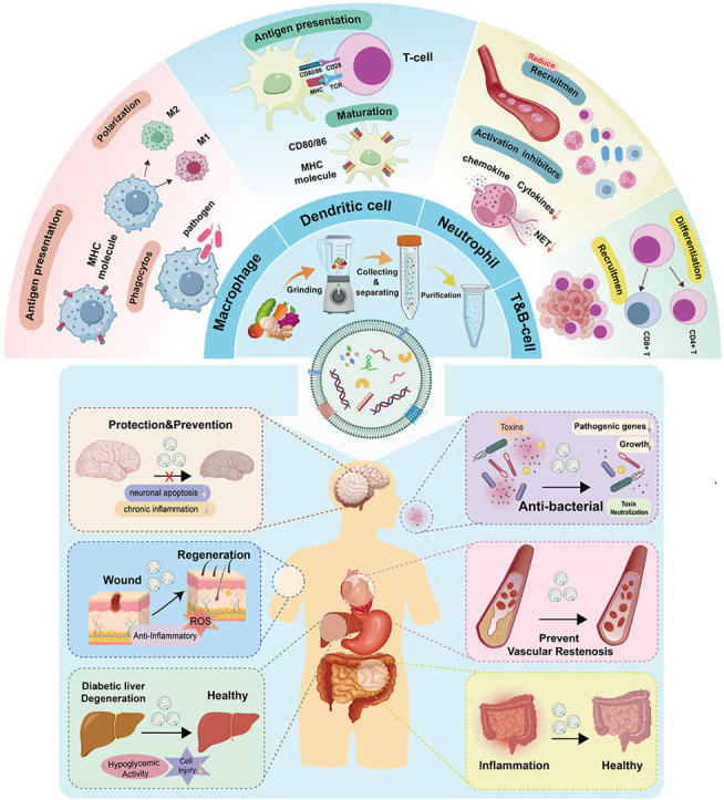
Immune dysregulation can result in sustained activation of the immune system, leading to systemic chronic inflammation. This condition significantly disrupts immune homeostasis and is intimately associated with the onset of numerous chronic systemic diseases. Currently, the treatment of diseases related to immune dysregulation confronts several challenges, most notably the substantial side effects and inconsistent efficacy of long-term immunosuppressive drug use. Consequently, developing immunomodulatory strategies that balance efficacy and safety has emerged as a prominent research focus. Plant-derived vesicle-like nanoparticles (PVLNs), natural nanomaterials secreted by plant cells, exhibit significant potential in immunomodulation owing to their excellent biocompatibility, minimal immunogenicity, and cross-species communication capabilities. This paper reviews the biogenesis, composition, and properties of PVLNs, emphasizing the mechanisms of innate and adaptive immunomodulation they mediate and their applications in diseases characterized by immune disorders. It also analyzes the challenges related to target delivery, stability optimization, drug loading, and storage encountered in their engineering. In the future, as the mechanisms of PVLNs are more deeply understood and nanotechnology continues to advance, their potential in precision immunotherapy and clinical translation is anticipated to be further augmented.

Keywords: Engineered modifications; Immunomodulation; Natural nanomaterials; Plant vesicles.


Conflict of interest statement

The authors declare no conflict of interest.

Figures

Image 1
Graphical abstract
Fig. 1
Fig. 1
Origin, composition, and biogenesis of PVLNs. PVLNs can be derived from various plants, and their components primarily include lipids, nucleic acids, proteins, and various plant-derived bioactive substances. There are roughly three pathways for PVLNs biogenesis: (1) MVB pathway: Early endosomes (ESE) are formed through encapsulation of extracellular components and membrane proteins via invagination of the plasma membrane. These endosomes mature into multivesicular bodies (MVBs), which then fuse with the plasma membrane to release vesicles into the extracellular space. (2) EXPO pathway: MVBs are formed after material exchange with the Golgi apparatus, and upon maturation, they fuse with the plasma membrane to release vesicles. (3) Vacuolar pathway: MVB-derived vesicles fuse with vacuolar vesicles to form intraluminal vesicles (ILVs), which are then transported to the extracellular space through fusion with the plasma membrane.
Fig. 2
Fig. 2
The mechanisms of PVLNs regulating the macrophage phagocytosis, polarization, and antigen presentation capabilities.
Fig. 3
Fig. 3
The pathway mechanism of PVLNs regulating dendritic cell differentiation.
Fig. 4
Fig. 4
The pathway mechanism of PVLNs in regulating the formation of neutrophil extracellular traps (NETs).
Fig. 5
Fig. 5
The mechanism of PVLNs in regulating the pathways of T cell activation, differentiation, recruitment, and reprogramming.
Fig. 6
Fig. 6
Administration routes and mechanisms of action of PVLNs in treating various immune-related diseases.
Fig. 7
Fig. 7
Application of Plant-Derived Exosome-Like Nanoparticles (PVLNs) in Digestive Diseases. (A) Diagram illustrating the extraction process of Ginger-Derived Exosome-Like Nanoparticles (GELNs). (B) Mechanisms of GELNs internalization by intestinal cells. Caco-2 cell nuclei were labeled with DAPI (blue), the cell membrane with DIO (green), and GELNs with PKH26 (red). GELNs uptake by small intestinal cells occurs via macropinocytosis and caveolin-mediated endocytosis. (C) Effects of GELNs on the expression of inflammation-related genes. Reprinted (adapted) with permission from (Yin L, Yan L, Yu Q et al. Characterization of the MicroRNA Profile of Ginger Exosome-like Nanoparticles and Their Anti-Inflammatory Effects in Intestinal Caco-2 Cells). Copyright © 2022, American Chemical Society. Licensed under the Creative Commons Attribution International License (CC BY 3.0).
Fig. 8
Fig. 8
Application of PVLNs in Treating Skin Wounds. (A) Schematic Illustration of the BFR-EVs Isolation Procedure: This section provides a visual representation of the steps involved in isolating BFR-EVs. (B) Scavenging Effect of BFR-EVs on Excess Intracellular ROS: Changes in intracellular ROS levels induced by H2O2 and UVB in human dermal fibroblasts (HDFs) treated with BFR-EVs were assessed using H2DCFDA staining and flow cytometric analysis. (C) BFR-EVs reduced the Expression Levels of Oxidative Stress-Related mRNA (MMP-1 and MMP-3) in UVB-induced HDFs: This reduction was quantified, demonstrating the potential of BFR-EVs in mitigating oxidative stress. (D) Scratch Closure Assay (Scale Bar = 200 μm) and 24-Hour Post-Transwell Migration Assay: CV staining was performed on HDFs that migrated to the bottom of the porous membrane, showing that BFR-EVs enhanced the proliferation and migration of HDFs. (E) BFR-EVs Inhibited Gene Expression of Pro-Inflammatory Cytokines in LPS-Stimulated RAW 264.7 Cells: This indicates the anti-inflammatory potential of BFR-EVs when applied to immune cells. Copyright © Antioxidants, 2023. Licensed under the Creative Commons Attribution International License (CC BY 4.0).
Fig. 9
Fig. 9
Applications of PVLNs in Metabolic Diseases. (A) Potential molecular signaling pathways of exosome-like nanoparticles from mung bean sprouts combating diabetic effects. (B) Morphology of MELNs and visualization by TEM. (C) Liver morphology changes and HE staining results. MELNs ameliorate liver pathology and reduce lipid accumulation. (D) In vivo expression of oxidative stress-related proteins. (E) GLUT4 expression at the cell membrane. MELNs enhance GLUT4 membrane translocation. (F) Protein expression of the PI3K/GSK-3β pathway (P-GSK-3β/GSK-3β) in liver tissue. Copyright © Springer Nature, 2023. Licensed under the Creative Commons Attribution International License (CC BY 4.0).
Fig. 10
Fig. 10
Application of PVLNs in cardiovascular disease. (A) A schematic representation of the mechanism through which exosome-like nanoparticles derived from Panax quinquefolium mitigate ischemia-reperfusion injury by modulating microglial polarization. (B) A schematic depiction of the isolation process for Panax-derived nanoparticles (PDNs). (C) Representative images of TTC staining in rats 72 h post-reperfusion, alongside quantitative analysis of infarct volumes in each group, demonstrate that PDNs attenuate ischemia-reperfusion injury in rats. (D) The proportions of M1 and M2 type microglia in the rat brain were measured 72 h following transient middle cerebral artery occlusion (tMCAO). (E) PDNs reduced microglia-induced inflammation following cerebral ischemia/reperfusion (CI/R), as evidenced by the concentrations of inflammatory cytokines TNF-α, IL-6, and IL-10 in brain tissues 72 h post-reperfusion. (F) Representative protein blot images and quantitative analyses of PI3k/Akt pathway-associated protein expressions illustrate that PDNs influence microglial polarization via the PI3k/Akt pathway. Copyright © J Nanobiotechnol, 2023. Under the terms of the Creative Commons Attribution International License (CC BY 4.0).
Fig. 11
Fig. 11
Application of PVLNs in Neurodegenerative Disease. (A) Characterization of LRM-ELNs, including TEM analysis, size measurement, and zeta potential assessment. (B) LRM-ELNs reduced Aβ-induced cytotoxicity and apoptosis in HT22 cells, as shown by MTT assay and flow cytometry. (C) Effects of LRM-ELNs on the mitochondrial apoptosis pathway in Aβ-induced HT22 cells. Protein levels of Bax/Bcl2 and Cleaved Caspase-3 were determined by Western blot. (D) Effects of LRM-ELNs on antioxidant indices in HT22 cells, including levels of ROS. (E) Effects of LRM-ELNs on the Nrf2/HO-1/NQO1 signaling pathway in Aβ-induced HT22 cells, analyzed via Western blot. Copyright © Food, 2024. (CC BY 4.0).
Fig. 12
Fig. 12
Application of PVLNs in Infectious diseases. (A) The overview diagram of GELNs inhibiting the pathogenicity of Porphyromonas gingivalis. (B)The uptake of GELNs and lipid nanoparticles (red) by Pseudomonas gingivalis (green). The uptake behavior of GELNs by Porphyromonas gingivalis depends on HBP35.(C)Hematoxylin staining of the oral section of control, P. gingivalis-infected mice, and GELN-treated mice. B, alveolar bone crest; PDL, periodontal ligament; E, epithelial cells. GELNs can reduce the cellular infiltration in the periodontal ligament area of mice infected with Pseudomonas gingivalis. (D)The wild type and HBP35 mutant of Porphyromonas gingivalis were co-incubated with GELNs for 24 h to determine bacterial growth through the optical density at 600 nm. (E)The expression of IL-1β and IL-6 mRNA in the oral tissues of mice was analyzed by RT-qPCR. (F)The attachment of Porphyromonas gingivalis to gingival epithelial cells (human telomerase-immortalized gingival keratinocytes [TIGKs]) with or without GELNs treatment under light focusing microscopy. (G)The distance from the cementoenamel junction (CEJ) to the alveolar bone crest (ABC) in the interdental space was measured linearly by MicroCT to reflect the alveolar bone loss in mice. copyright iScience, 2019. Under the terms of the Creative Commons Attribution International License(CC BY 4.0).

References

    1. Furman D., Campisi J., Verdin E., Carrera-Bastos P., Targ S., Franceschi C., Ferrucci L., Gilroy D.W., Fasano A., Miller G.W., Miller A.H., Mantovani A., Weyand C.M., Barzilai N., Goronzy J.J., Rando T.A., Effros R.B., Lucia A., Kleinstreuer N., Slavich G.M. Chronic inflammation in the etiology of disease across the life span. Nat Med. 2019;25:1822–1832. doi: 10.1038/s41591-019-0675-0. - DOI - PMC - PubMed
    1. Hegarty L.M., Jones G.-R., Bain C.C. Macrophages in intestinal homeostasis and inflammatory bowel disease. Nat. Rev. Gastroenterol. Hepatol. 2023;20:538–553. doi: 10.1038/s41575-023-00769-0. - DOI - PubMed
    1. Ridker P.M. Anticytokine agents: targeting interleukin signaling pathways for the treatment of atherothrombosis. Circ. Res. 2019;124:437–450. doi: 10.1161/CIRCRESAHA.118.313129. - DOI - PMC - PubMed
    1. King G.L. The role of inflammatory cytokines in diabetes and its complications. J. Periodontol. 2008;79:1527–1534. doi: 10.1902/jop.2008.080246. - DOI - PubMed
    1. Ziogas A., Bruno M., van der Meel R., Mulder W.J.M., Netea M.G. Trained immunity: target for prophylaxis and therapy. Cell Host Microbe. 2023;31:1776–1791. doi: 10.1016/j.chom.2023.10.015. - DOI - PubMed