. 2025 Oct 2;23(1):408.
doi: 10.1186/s12964-025-02414-6.

IGFBP2 alteration contributes to prostate cancer progression by modulating prostate stroma activation

Affiliations

IGFBP2 alteration contributes to prostate cancer progression by modulating prostate stroma activation

Mingguo Huang et al. Cell Commun Signal. .

Abstract

Background: Insulin-like growth factor (IGF) binding protein-2 (IGFBP2) is a secretory protein that modulate the activity of IGFs. It is highly expressed in various cancers such as prostate cancer (PCa), in which it may play a controversial role in tumor progression, however, the molecular mechanisms of IGFBP2 in PCa progression remain unclear.

Methods: In this study, we examined the expression pattern and role of IGFBP2 in PCa cells and the stroma during prostate tumor cell progression.

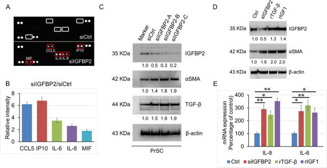
Results: IGFBP2 was highly expressed in LNCaP cells and prostate stromal fibroblasts (PrSC) and was mainly secreted by PrSCs. Tumor cell growth and invasiveness were not directly affected by treatment with IGFBP2 siRNAs (siIGFBP2) or recombinant IGFBP2 (rIGFBP2). However, decreased expression of IGFBP2 significantly increased PrSC activation and the secretion of pro-tumorigenic cytokines IL-6, IL-8, IP10, and CCL5 through upregulation of TGF-β, which subsequently enhanced prostate tumor cell progression. Clinically, low expression of stromal IGFBP2 was associated with a high reactive prostate stroma, advanced PCa progression, and increased IGFBP2 levels in the serum.

Conclusion: Here, we provide mechanistic evidence that IGFBP2 act as a critical regulatory factor in the activation of prostate stromal microenvironment and contributes to aggressive PCa progression.

Keywords: IGFBP2; Insulin-like growth factor binding protein-2; Prostate cancer; Prostate stromal cells.


Conflict of interest statement

Declarations. Ethics approval and consent to participate: The Institutional Review Board of the Akita University School of Medicine approved all the experiments, which were performed after obtaining individual written informed consent. Consent for publication: All Authors agreed to the manuscript. Competing interests: The authors declare no competing interests. The authors hereby certify and declare that all authors participated in the study and that all authors have seen and approved the manuscript. I also certify that the contents of this manuscript are not now under consideration for publication elsewhere, and the contents will not be copyrighted, submitted, or published elsewhere while acceptance by the Journal is under consideration.

Figures

Fig. 1
Fig. 1
Conditioned Media from PrSC with reduced IGFBP2 both promote PCa cell growth and invasiveness. A and (B) 1 × 105 of LNCaP, PC3, DU145, and prostate stromal cells (PrSC) were cultured in a 35-mm dish with DMEM containing 2% FBS without antibiotics for 24 h. The cells and conditioned medium (CM) were harvested (A) Proteins from equivalent volumes of lysates and CM was subjected to western blot analysis with anti-human IGFBP2 antibody (B) The IGFBP2 concentration of the CM derived from the cells was measured using a human IGFBP2 ELISA Kit (C) PCa cells were cultured in DMEM containing 2% FBS with 100 ng/ml rIGFBP2 or 100 nM siIGFBP2 or CM from PrSC previously cultured in the presence or absence of 100 nM siIGFBP2 for 24 h. The MTT assay was performed, and cell viability was compared with that of cells cultured with FBS. *P < 0.05, **P < 0.01 (D) PCa cells cultured for 24 h in DMEM containing 2% FBS with 100 ng/ml rIGFBP2 or 100 nM siIGFBP2, or CM from PrSC previously cultured in the presence or absence of 100 nM siIGFBP2 for 24 h. Invaded cells were counted and their numbers were compared with those of cells cultured with FBS. *P < 0.05, **P < 0.01
Fig. 2
Fig. 2
Decreased IGFBP2 enhances PrSC activation and pro-tumorigenic cytokine secretion. A and (B) 1 × 105 PrSC were cultured in a 35-mm dish with 100 nM of siIGFBP2 or siCtrl for 24 h. The CM was collected, and serum cytokine levels analyzed using the comprehensive proteome profiler human cytokine array kit (A) The signal intensity in the array images was quantified and scored using Densitograph software. The signals for CCL5, IP10, IL-6, IL-8, and MIF in the array images are indicated by rectangular boxes (B) The fold changes among samples were analyzed and compared by R&D Systems proteome profiler array analysis (C) PrSC were treated with several IGFBP2 RNAs (siIGFBP2-A, B, and C) for 24 h. Equivalent amounts of protein (10 µg) from the cells were analyzed using anti-IGFBP2, anti-αSMA, anti-TGF-β, and anti-β-actin antibodies (D) and (E) PrSC were treated with 100nM siIGFBP2, 10 ng/ml rTGF-β, or 10 ng/ml rIGF1 for 12 h. Protein from the cultured cells was evaluated by western blot with anti-IGFBP2 anti-αSMA antibodies (D). Total RNA was extracted from the cells, and IL-8 and IL-6 mRNA levels were measured by quantitative reverse transcription PCR and compared with those of untreated cells (E). *P < 0.05, **P < 0.01
Fig. 3
Fig. 3
Decreased IGFBP2 enhances PrSC activation and increases PCa progression in vivo PC-3 M-luc-C6 cells were intraperitoneally injected into mice with PrSC cells previously treated with 100 nM siCtrl or siIGFBP2 for 24 h (Ctrl and siIGFBP2 groups, three mice per group) (A) At 12 days inoculation, bioluminescence was used to detect intraperitoneal tumor cell expansion by the intraperitoneal injection of luciferin (B) The level of luciferase activity was significantly higher in the siIGFBP2 group compared with the Ctrl group (C) and (D) Slides of mouse tumor tissues were subjected immunohistochemistry to detect the expression of IGFBP2 and αSMA, and the expression levels of IGFBP2 and αSMA were scored on a semiquantitative scale. bar, 100 μm. ns: not significant, *P < 0.05
Fig. 4
Fig. 4
Low expression of stromal IGFBP2 is correlated with prostate stroma activation and advanced PCa progression. A Immunohistology staining of tissue samples from 168 patients with PCa radical prostatectomy using anti-human αSMA and IGFBP2 antibodies, respectively. The percentage of αSMA-positive stromal cells surrounding of cancer cells was calculated, and the staining level was determined by the staining percentage score characterized as low (≤ 5%, n = 65), moderate (5–20%, n = 71), or high (> 20%, n = 31). The intensity of IGFBP2-stained stromal cells in the area surrounding the cancer cells was evaluated, and the staining index was determined by the staining intensity score (B) The relationship between αSMA and IGFBP2 expression was evaluated. The IGFBP2 staining level in the patient group with low αSMA staining was significantly higher compared with that of the patient groups with medium or high αSMA staining, respectively. The IGFBP2 staining level was significantly higher in the patient group with medium αSMA staining compared with that of the patient group with high αSMA staining. *P < 0.05, **P < 0.01 (C) Serum IGFBP2 levels from 40 controls and 168 patients with PCa were measured using a human IGFBP2 ELISA Kit. Serum IGFBP2 levels were significantly higher in PCa patients compared with those in the controls (71.3 ± 17.0 and 191.4 ± 13.5 ng/ml, respectively). Serum IGFBP2 levels were significantly higher in the PCa patient group with high αSMA staining compared with those in the patient groups with medium or low αSMA staining (137.6 ± 22.1, 186.6 ± 41.8, and 215.9 ± 41.3 ng/ml, respectively). * P < 0.05, **P < 0.01 (D) The relationship between αSMA expression level and pathological T stage (pT) was evaluated. Patients with low αSMA staining comprised 59,1% of pT1, 34.8% of pT2, and 6.1% of pT3 or 4. Patients with medium αSMA staining comprised 36.6% of pT1, 39.4% of pT2, and 24.0% of pT3 or 4. Patients with high αSMA staining comprised 25.8% of pT1, 45.1% of pT2, and 29.1% of pT3 or 4. *P < 0.05, **P < 0.01
Fig. 5
Fig. 5
Schematic role of IGFBP2 in prostate stromal cell activation and tumor progression. IGFBP2 secretion was increased in activated PrSC, which was affected by growth and stimulatory factors. Activated PrSC promotes PCa tumor progression through the production of pro-tumorigenic cytokines, such as IL-6, IL-8, IP10, and CCL5. Decreased expression and secretion of IGFBP2 may be implicated in the regulation of prostate stromal cell activation and PCa progression

References

    1. Jemal A, Siegel R, Ward E, Murray T, Xu J, Thun MJ. Cancer statistics, 2007. CA Cancer J Clin. 2007;57:43–66. - PubMed
    1. Ito K. Prostate cancer in Asian men. Nat Rev Urol. 2014;11:197–212. - PubMed
    1. Kogan-Sakin I, Cohen M, Paland N, Madar S, Solomon H, Molchadsky A, Brosh R, Buganim Y, Goldfinger N, Klocker H, et al. Prostate stromal cells produce CXCL-1, CXCL-2, CXCL-3 and IL-8 in response to epithelia-secreted IL-1. Carcinogenesis. 2009;30:698–705. - PubMed
    1. Ao M, Franco OE, Park D, Raman D, Williams K, Hayward SW. Cross-talk between paracrine-acting cytokine and chemokine pathways promotes malignancy in benign human prostatic epithelium. Cancer Res. 2007;67:4244–53. - PubMed
    1. Pollak MN, Schernhammer ES, Hankinson SE. Insulin-like growth factors and neoplasia. Nat Rev Cancer. 2004;4:505–18. - PubMed

MeSH terms

Substances

LinkOut - more resources