. 2025 Oct 3;11(1):286.
doi: 10.1038/s41531-025-01136-3.

Evaluating Parkinson's disease biomarkers in substantia nigra following sublethal γ-radiation exposure in a large animal model

Affiliations

Evaluating Parkinson's disease biomarkers in substantia nigra following sublethal γ-radiation exposure in a large animal model

Erin K Murphy et al. NPJ Parkinsons Dis. .

Abstract

Idiopathic Parkinson's Disease (iPD) involves genetic and environmental factors, including ionizing radiation. While high-dose radiation induces neurodegeneration, the effects of low-dose radiation (LDR) remain unclear. This study examined the impact of a single acute total-body LDR exposure (1.79 Gy) on the substantia nigra (SN) of swine, a large mammal model closely resembling humans. Fourteen male Göttingen minipigs were assigned to radiation (RAD; n = 6) or sham (SH; n = 8) groups. We analyzed iPD-related markers (α-synuclein, phosphorylated α-syn, tyrosine hydroxylase), genetic PD markers (LRRK2, GBA, VPS13C, Cathepsin D), neuroinflammation (GFAP), and mitochondrial proteins (ATP5A, SDHB, NDUF8). No significant molecular, histological, or immunohistochemical differences were observed between RAD and SH animals. LRRK2 was undetectable, and no structural damage or neuroglial changes were found. These findings suggest that single acute LDR exposure does not elicit short-term PD-related alterations in the SN of swine, although long-term or cumulative effects warrant further investigation.


Conflict of interest statement

Competing interests: The authors declare no competing interests.

Figures
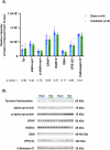
Fig. 1
Fig. 1. Single exposure to total body radiation did not alter Parkinson’s related proteins in the substantia nigra.
A Western blot densitometric analysis for PD related markers in the substantia nigra from the brains of Sham (n = 8) (blue) and Irradiated (n = 6) (green) Gottingen minipigs 33–35 days post total body radiation. B Representative western blots for all proteins are shown. Each blot was run in triplicate, and the graphs represent the average of 3 runs. * indicates p < 0.05 as determined by unpaired 2-tailed t-test. Error bars represent the standard error of the mean. Original full-length blots are presented in Supplementary Figs. 3–4.
Fig. 2
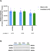
Fig. 2. Single exposure to total body radiation did not alter mitochondrial related proteins in the substantia nigra.
A Western blot densitometric analysis for mitochondrial related markers in the substantia nigra from the brains of Sham (n = 8) (blue) and Irradiated (n = 6) (green) Gottingen minipigs 33–35 days post total body radiation. B Representative western blots for all proteins are shown. Each blot was run in triplicate, and the graphs represent the average of 3 runs. * indicates p < 0.05 as determined by unpaired 2-tailed t-test. Error bars represent the standard error of the mean. Original full-length blots are presented in Supplementary Fig. 5.
Fig. 3
Fig. 3. Histological and immunohistochemical outcomes in the substantia nigra of Gottingen minipigs after total body radiation.
The figure shows various histological and IHC stains for the SN of RAD and SH swine. HE stain (panels A and E) of the SN region in RAD vs. SH did not demonstrate any obvious difference between the two conditions. No vascular or perivascular lesions were observed. Also, to notice, the absence of neuromelanin pigment in the dopaminergic neurons of the swine SN. CV stain (panels B and F) did not evidence cellular or subcellular morphometric changes in RAD vs. SH SN cells, either neuronal cells or glial cells. The TH stain (panels C and G) clearly show positivity for tyrosine hydroxylase (TH) of the dopaminergic neurons of the swine SN. No difference in terms of IHC intensity between RAD vs. SH condition were detectable. GFAP stain (panels D and H) shows the absence of significant differences in terms of astroglial reactivity between RAD and SH in the SN of swine. IHC immunohistochemistry; HE hematoxylin and eosin; CV cresyl violet; TH tyrosine hydroxylase; GFAP glial fibrillary acidic protein; RAD irradiated animals; SH sham, non-irradiated animals; SN substantia nigra. The bottom left line underneath panel E. indicates the corresponding length (200 μm) of all images taken at the same level of magnification.

References

    1. Pringsheim, T., Jette, N., Frolkis, A. & Steeves, T. D. The prevalence of Parkinson’s disease: a systematic review and meta-analysis. Mov. Disord.29, 1583–1590 (2014). - PubMed
    1. Ben-Shlomo, Y. et al. The epidemiology of Parkinson’s disease. Lancet403, 283–292 (2024). - PMC - PubMed
    1. Ball, N., Teo, W. P., Chandra, S. & Chapman, J. Parkinson’s disease and the environment. Front. Neurol.10, 218 (2019). - PMC - PubMed
    1. Tsalenchuk, M., Gentleman, S. M. & Marzi, S. J. Linking environmental risk factors with epigenetic mechanisms in Parkinson’s disease. NPJ Parkinsons Dis.9, 123 (2023). - PMC - PubMed
    1. Tansey, M. G. et al. Inflammation and immune dysfunction in Parkinson disease. Nat. Rev. Immunol.22, 657–673 (2022). - PMC - PubMed

LinkOut - more resources