. 2025 Nov;10(11):2736-2748.
doi: 10.1038/s41564-025-02135-z. Epub 2025 Oct 3.

Superinfection promotes replication and diversification of defective HIV-1 proviruses in people with non-suppressible viraemia

Affiliations

Superinfection promotes replication and diversification of defective HIV-1 proviruses in people with non-suppressible viraemia

Vivek Hariharan et al. Nat Microbiol. 2025 Nov.

Abstract

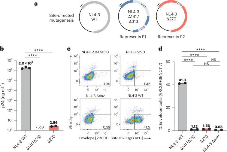
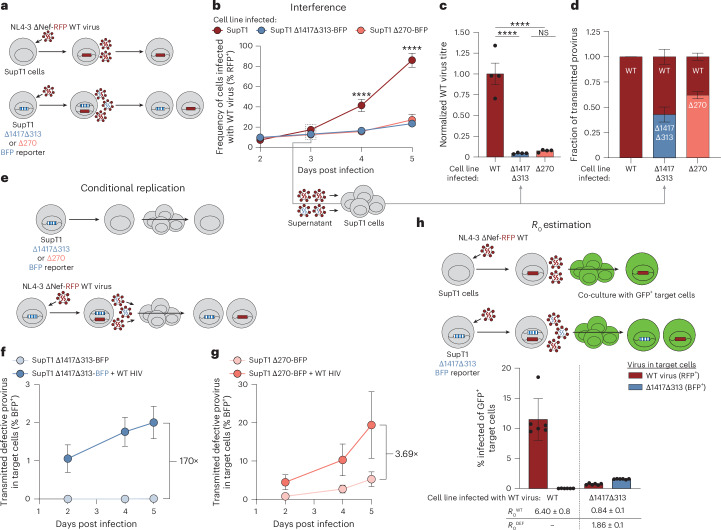
During replication of some RNA viruses, defective particles can spontaneously arise and interfere with wild-type (WT) virus replication. However, these defective interfering particles (DIPs) have not been reported in people with HIV-1 (PWH). Here we find DIPs in PWH who have a rare, polyclonal form of non-suppressible viraemia (NSV). We characterized the source of NSV in two PWH who never reached undetectable viral load despite adherence to antiretroviral therapy (ART). Remarkably, in each participant, we found a diverse set of defective viral genomes sharing the same fatal deletions. This paradoxical accumulation of mutations by viruses with fatal defects was driven by superinfection with intact viruses, resulting in mobilization of defective genomes and accumulation of additional mutations during untreated infection. These defective proviruses interfere with WT virus replication, conditionally replicate and, in one case, have an R0 > 1, enabling in vivo spread. Despite this, clinical outcomes showed no beneficial effect of these DIPs. These findings demonstrate that fatally defective proviruses, traditionally considered evolutionary dead ends, can replicate and diversify upon superinfection without preventing disease progression.


Conflict of interest statement

Competing interests: R.F.S. is an inventor on a patent application for the intact proviral DNA assay (IPDA) (patent no. PCT/US16/28822) filed by Johns Hopkins University and licensed by AccelevirDx. F.R.S. received payments from Gilead Sciences for participating at scientific meetings. The other authors declare no competing interests.

Figures

Fig. 1
Fig. 1. Failure to suppress viraemia is characterized by diverse virus in plasma and high infected-cell frequency.
a,b, Plasma HIV-1 RNA and CD4+ T cell counts over time for P1 (a) and P2 (b). Numbers above squares represent CD4+ T cell percentages. The dotted line at 20 copies per ml represents the current limit of detection for the clinical HIV-1 viral load assay. Data are for periods with optimal adherence. For P2, there was a preceding period of suboptimal adherence (see text and Extended Data Fig. 1a). c,d, Neighbour-joining phylogenetic trees of p6-RT single genome sequences obtained from plasma viral RNA for P1 (c) and P2 (d). Phylogenetic tree tip labels are colour coded according to the plasma collection timepoint in a. Phylogenetic trees are rooted to HXB2, and HIV-1 coordinates refer to the HXB2 reference genome. Tree nodes with bootstrap values >80 are marked with asterisks. e, Intact proviral DNA frequencies as measured by the IPDA. The percentage of proviruses classified as intact by the IPDA is shown on the top. Asterisks represent analyses performed on total white blood cells that were corrected on the basis of the CD4+ T cell percentage at the time of sampling. f, Infectious units per million (IUPM) CD4+ T cells as measured by the quantitative viral outgrowth assay. g, IUPM to IPDA intact ratio as measured by dividing the IUPM by the closest IPDA timepoint value. TAF, tenofovir alafenamide; FTC, emtricitabine; BIC, bictegravir; DRV/c, darunavir/cobicistat; b.i.d., bis in die (twice daily); DRV/r, darunavir/ritonavir; DTG, dolutegravir. Source data
Fig. 2
Fig. 2. Near full-length sequencing reveals dominant proviral populations with unique deletion signatures and diverse mutations across the viral genome.
a,e, Left: neighbour-joining phylogenetic trees of near full-length proviral sequences obtained by single genome sequencing from primary CD4+ T cells, rooted to HXB2. The colour of each branch tip indicates sampling time as in Fig. 1a,b. Dashed branches indicate sequences with hypermutation. Right: highlighter plot with black lines representing nucleotide changes compared to the top sequence. Grey vertical bars represent deletions compared to HXB2. Highlighted areas represent recurrent deletion patterns of 1,417 nt, 313 nt (P1) and 270 nt (P2). IPDA primer probe regions are highlighted at the top. Green arrowheads point to sequences with significant G→A hypermutation. b,f, Mapped sequences of prominent deletion signatures found in majority of proviruses in P1 and P2 compared to HXB2. c,g, Dot plots representing the number of mutations between each near full-length sequence and the majority consensus sequences. Data represent mean ± s.d. (c) n = 32 and (g) n = 65. d,h, Histogram (bin width of 5) representing the nucleotide differences in unique proviruses from each participant. Dashed line represents the mean number of nucleotide differences between all unique proviral sequences.
Fig. 3
Fig. 3. Defective proviruses dominate the proviral landscape and contribute to NSV.
a, Location of primers (arrows) and probes (rectangles with vertical bars) to specifically quantify deletion signatures in viral RNA and DNA. Probes span the deletion region. b, Representative two-dimension plots of dPCR showing duplex amplification of intact proviruses and proviruses of interest by deletion-specific assays. c, Longitudinal quantification of proviruses with specific deletions of interest. Data are mean of technical triplicates (n = 3). Asterisks represent analyses performed on total white blood cells that were corrected on the basis of the CD4+ T cell percentage at the time of sampling. d, CD4+ T cells from P1 and P2 were cultured for 48 or 72 h in the presence of emtricitabine (FTC), tenofovir disoproxil fumarate (TDF), dolutegravir (DTG) and anti-CD3/CD28 beads. The virion-associated RNA in the supernatant was measured by RT–dPCR. Error bars represent s.e.m. Pie chart shows percentage of HIV-1 RNA copies with deletion normalized to the total number of copies. e, Plasma virion-associated RNA was measured by RT–dPCR. Data are mean ± s.e.m. performed in at least technical triplicates (n = 3–8). Pie chart shows percentage of HIV-1 RNA copies with deletion normalized to the total number of copies. For P1, total proviruses were quantified by measuring the highly conserved region LTR-gag. For P2, proviruses without the 270-nt deletion were measured using a primer–probe set inside of the deletion (WT-270). Source data
Fig. 4
Fig. 4. Deletions found in proviruses abolish virus production in vitro.
a, Prominent deletions found in proviruses were introduced into an NL4-3 expression plasmid by site-directed mutagenesis. Arrows represent orientation of the provirus within the expression plasmid. b, Virus produced upon HEK293T transfection was pelleted by ultracentrifugation, and p24 was measured by ELISA. Lower limit of detection (LoD) was 0.625 ng ml−1. Data represent mean ± s.d. c, Representative flow cytometry plots of the surface staining of transfected HEK293T cells with viability dye and bNAbs (VRC01 and 3BNC117). d, Surface staining of HIV-1 Env with bNAbs (VRC01 and 3BNC117) on HEK293T cells 24 h after transfection. Data represent mean ± s.e.m. (b,d) Statistical significance between conditions was determined using one-way analysis of variance (ANOVA). ****P < 0.0001, NSP > 0.01. Source data
Fig. 5
Fig. 5. Cell culture model to assess superinfection in vitro.
a, Schematic for assessment of superinfection in vitro. Top: SupT1 cells are infected with NL4-3-ΔNef-RFP WT virus. Bottom: model cell lines transduced with reporter defective viruses representing P1 (Δ1417Δ313) or P2 (Δ270) are infected with NL4-3-ΔNef-RFP WT virus. Virions produced by these superinfected cells may package RNA from the defective (blue) or WT (red) viral genome. b, Frequency of cells infected with the WT virus (% RFP+) measured over time. Data represent mean ± s.d. c, Viral supernatant from day 3 of infection of each cell line was used to infect target SupT1 cells to determine the viral titre of WT virus after a single round of infection. WT viral titre was normalized to virus from the WT SupT1 cell line. Data represent mean ± s.e.m. d, Viral supernatant from day 3 of infection of each cell line was used to infect SupT1 cells to determine the fraction of transmitted provirus after a single round of infection as measured by flow cytometry. e, Schema for assessment of conditional replication in vitro. Model cell harbouring reporter defective viruses are either mock infected or infected with WT virus. After 3 days of culture, the resulting supernatant is used to infect target SupT1 cells. f,g, Frequency of target SupT1 cells infected with defective provirus measured over time. Data represent mean ± s.d. h, Top: schema for three-colour experiment to estimate R0 in vitro. WT SupT1 or SupT1-Δ1417Δ313-BFP are infected with WT virus. After 2 days, the cells were co-cultured with excess GFP+ SupT1 target cells. Bottom: frequency of target GFP+ SupT1 target cells infected with either WT virus (red) or defective virus (blue). R0 values ± s.e.m. for both the WT virus (R0WT) and defective virus (R0DEF) are listed at the bottom. Data represent mean ± s.d. (bd) n = 4 for all conditions assessed. (fh) n = 6 for all conditions assessed. (b,c) Statistical significance between cell lines was determined using one-way ANOVA. ****P < 0.0001, NSP > 0.05. Source data
Fig. 6
Fig. 6. Single p24+ cell sequencing identifies dually infected cells in vivo.
a, Experimental design to characterize p24+ cells and detect multiple integrated proviruses. Sorted p24+ cells are lysed and spread over multiple wells. Near full-length proviral amplification, agarose gel electrophoresis and sequencing are conducted on the cell lysate. Example agarose gel electrophoresis is shown. b,c, Neighbour-joining phylogenetic tree of near full-length proviral genomes from p24+ cells for P1 (b) and P2 (c). Highlighter plot with black lines represent nucleotide changes compared to the top sequence. Grey boxes represent deletions compared to the top sequence of each tree. Tree nodes with bootstrap values >80 are marked by asterisks. 5′ internal deletions are boxed in black and annotated. Common deletions found in P1 and P2 are boxed and annotated. Phylogenetic tree tips with grey boxes represent single provirus found from a sorted cell. Coloured boxes represent multiple proviruses found from a single sorted cell, probably representing multiple integrated proviruses within one cell. Coloured arrowheads point to multiple proviruses found in a single sorted cell.
Extended Data Fig. 1
Extended Data Fig. 1. Extended clinical history of P2 and phylogenetic analyses.
(a) Extended clinical history for P2. (Top) Prescribed ART regimen for P2. (Middle) Plasma HIV-1 RNA and CD4+ T cell counts for P2. Gray shaded area represents time prior to optimal adherence of ART. Gray viral load circles indicate values below the limit of quantification. (Bottom) Plasma HIV-1 RNA and CD4+ T cell count for P2 for years on ART with optimal adherence (also shown in Fig. 1b). Phylogenetic tree tip labels are colour coded according to the plasma collection timepoint in Fig. 1b. (b) Neighbour-joining phylogenetic trees of env single genome sequences obtained from plasma-associated virions for P2. Tree nodes with bootstrap values above 80 are marked with asterisks. (c) Plots of root-to-tip distance and average pairwise distance of P6-RT plasma virus sequences for P1 and P2 showing lack of viral evolution during ART. Box and whisker plots with center lines indicating the median, boxes representing the interquartile range (IQR), and whiskers representing max to min values. Statistical significance calculated by ordinary one-way ANOVA. n.s.P > 0.05. n = 134 sequences analyzed for P1; n = 51 sequences analyzed for P2. 3TC: lamivudine; TDF: tenofovir disoproxil fumarate; TDF/3TC: tenofovir disoproxil fumarate/lamivudine; TAD/FTC: tenofovir alafenamide/emtricitabine; TDF/FTC/EFV: tenofovir disoproxil fumarate/emtricitabine/efavirenz; ETV: entecavir; TDF/FTC/RPV: tenofovir disoproxil fumarate/emtricitabine/rilpivirine; ATV: atazanavir; RTV: ritonavir; DRV: darunavir; RAL: raltegravir; DTG: dolutegravir. Source data
Extended Data Fig. 2
Extended Data Fig. 2. Validation of digital PCR assays and longitudinal quantification of defective proviruses during periods of suboptimal ART.
(a) Results of dPCR reaction using primer and probe sets to specifically amplify deletion regions of interest in 4 PWH on ART (040, 012, 017, and 274), P1, and P2. Horizontal bars represent mean and error bars represent SD, with each experiment performed in technical triplicate (n = 3). (b) Extended clinical history of P2 from Extended Data Fig. 1a (c) Longitudinal quantification of proviruses with specific deletion of interest using deletion-specific dPCR assay outlined in Fig. 3. WBCs: white blood cells (d) Longitudinal quantification of plasma virus with specific deletion of interest. Source data
Extended Data Fig. 3
Extended Data Fig. 3. QVOA wells with exponential outgrowth contain proviruses with and without the recurrent deletions.
(a and b) (Left) Neighbour-joining phylogenetic trees of single genome sequences obtained from outgrowth wells. Coloured boxes represent multiple sequences obtained from the same outgrowth well. The sequenced amplicon coordinates refer to the reference genome HXB2. Tree nodes with bootstrap values above 80 are marked with asterisks. (Right) Highlighter plot with black lines representing nucleotide changes compared to the top sequence of each tree. Gray vertical bars represent deletions compared to HXB2. Boxed regions represent recurrent deletion patterns of 1417 nt, 313 nt (P1), and 270 nt (P2).
Extended Data Fig. 4
Extended Data Fig. 4. Extended analysis of transfected HEK293T cells.
(a) Gating scheme used in flow cytometry to identify transfected cells expressing Env on the cell surface. A representative well of HEK293T cells transfected with NL4-3 WT and stained using a bNAb and secondary α-human IgG is shown here. (be) Surface staining of HIV-1 Env on HEK293T cells 24 h after transfection with various bNAbs (10E8v4, PGT121, PGT128, and PGDM1400). Horizontal bars represent mean and error bars represent SD, with each experiment performed in technical duplicates (n = 2). Statistical significance calculated by ordinary one-way ANOVA. **** P < 0.0001. Source data
Extended Data Fig. 5
Extended Data Fig. 5. Extended analysis of in vitro superinfection experiments.
(a) (Top) Construct map of NL4-3-ΔNef-BFP with the deletions marked. (Bottom) Flow cytometry gating of BFP+ sorted cells. (b) (Left) Schema of in vitro superinfection interference assay. (Right) Representative flow cytometry gating for each condition. (c) Comparison of the construct maps showing recurrent deletions identified in P1 and P2 from this study alongside engineered deletions in pNL4-3 from Pitchai et al. Light boxes represent deletion regions. The dark black square represents the central polypurine tract (cPPT). (d) (Top) Schema of the in vitro R0 estimation assay. (Bottom) Representative flow cytometry gating for each condition for the estimation of R0.
Extended Data Fig. 6
Extended Data Fig. 6. Single cell sorting validation.
(a) The probability of separating two genomes, if present, into distinct wells increases as the number of wells over which the cell lysate is distributed grows. (b) Live, single, CD4+ cells from a male uninfected donor were sorted into lysis buffer and distributed over 6 wells. A region of the sex-determining region Y gene was amplified, and the resulting gel shows only one band from each of the 6 sorted cells. Each red box represents the lysate from one cell spread over 6 PCR wells. A no-template control (water only) and positive control (DNA from an uninfected male) are included. n = 6 cells sorted and analyzed. (c) Uninfected donor CD4 + T cells were mixed with ACH-2 cells, stained for p24, and single-cell sorted. ACH-2 cells were lysed and distributed across 12 wells for near-full-length proviral amplification. The resulting agarose gel shows 12 wells per cell lysate (boxed). n = 6 cells sorted and analyzed. (d) Neighbor-joining phylogenetic tree from sorted, sequenced p24+ cells from a PWH with low-level viraemia. Gray boxes indicate a single provirus found in a sorted cell. Nodes with bootstrap values > 80 are marked with asterisks. Source data

Update of

References

    1. Chun, T.-W. et al. In vivo fate of HIV-1-infected T cells: quantitative analysis of the transition to stable latency. Nat. Med.1, 1284–1290 (1995). - PubMed
    1. Chun, T.-W. et al. Quantification of latent tissue reservoirs and total body viral load in HIV-1 infection. Nature387, 183–188 (1997). - PubMed
    1. Finzi, D. et al. Identification of a reservoir for HIV-1 in patients on highly active antiretroviral therapy. Science278, 1295–1300 (1997). - PubMed
    1. Wong, J. K. et al. Recovery of replication-competent HIV despite prolonged suppression of plasma viremia. Science278, 1291–1295 (1997). - PubMed
    1. Chun, T.-W. et al. Presence of an inducible HIV-1 latent reservoir during highly active antiretroviral therapy. Proc. Natl Acad. Sci. USA94, 13193–13197 (1997). - PMC - PubMed

LinkOut - more resources