. 2025 Oct 6;20(1):161.
doi: 10.1186/s13020-025-01218-7.

Electroacupuncture attenuates intestinal epithelial ferroptosis in inflammatory bowel disease via Piezo1-mediated mitochondrial homeostasis

Affiliations

Electroacupuncture attenuates intestinal epithelial ferroptosis in inflammatory bowel disease via Piezo1-mediated mitochondrial homeostasis

Haolong He et al. Chin Med. .

Abstract

Background: Inflammatory bowel disease (IBD) involves pathological mechanical forces transduced by mechanosensitive Piezo1 channels. While electroacupuncture (EA) alleviates IBD injury, its relationship with Piezo1-mediated ferroptosis remains unknown.

Methods: Dextran sulfate sodium (DSS)-induced IBD mice and mechanically stressed HIEC-6 intestinal epithelial cells received EA or pharmacological modulators. Pathological scoring, transmission electron microscopy (TEM), inflammatory cytokine assays, Western blotting, and immunofluorescence evaluated mitochondrial dynamics and ferroptosis markers to elucidate the Piezo1-ferroptosis axis and EA's regulatory role.

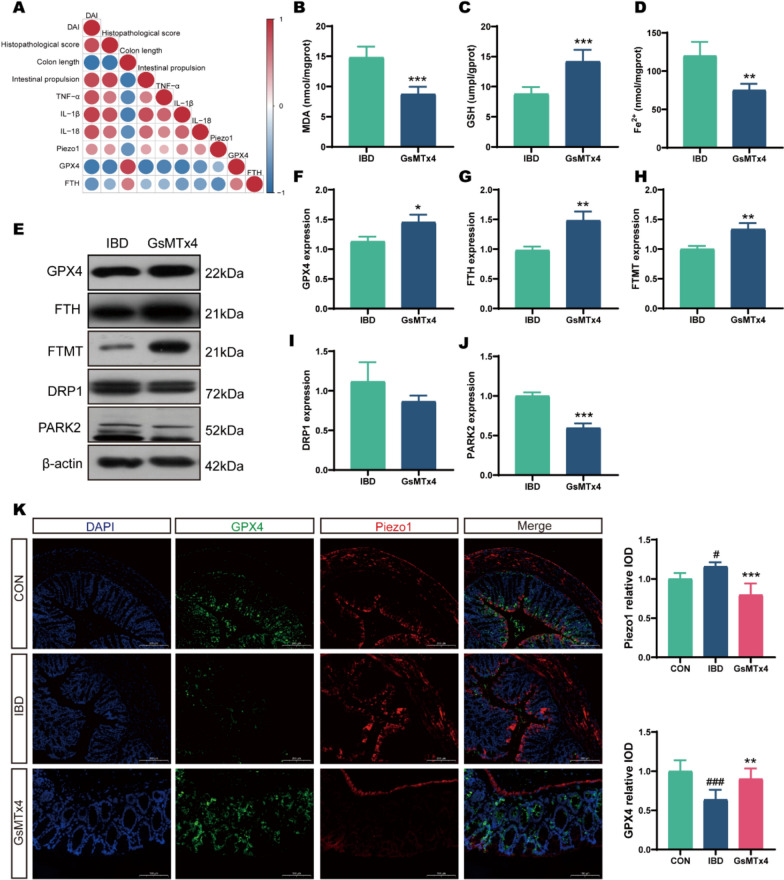
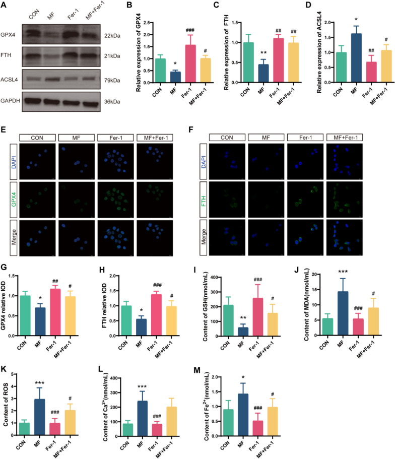
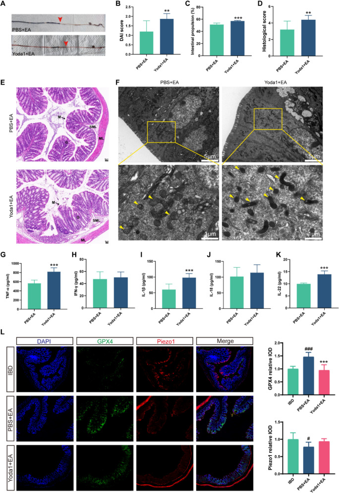
Results: EA significantly reduced disease activity index (DAI), histopathological scores, colon shortening, and pro-inflammatory cytokines in IBD mice. By inhibiting fission, indicated by a decrease in dynamin-related protein 1 (DRP1), and mitophagy, shown by a reduction in Parkinson protein 2 (PARK2), EA maintained mitochondrial homeostasis. This effect was similar to ferroptosis inhibitor ferrostatin-1 (Fer-1). Moreover, EA lessened RSL3-induced exacerbation of ferroptosis. In vitro, mechanical stress or the Piezo1 agonist Yoda1 induced ferroptosis, which was evident from increased acyl-CoA synthetase Long-chain family member 4 (ACSL4), reactive oxygen species (ROS), malondialdehyde (MDA) and Fe2⁺ levels, while decreased glutathione peroxidase 4 (GPX4), ferritin (FTH) and glutathione (GSH) levels. Critically, EA inhibited Piezo1 activation and counteracted Yoda1-aggravated epithelial ferroptosis in vivo.

Conclusion: Piezo1-mediated mitochondrial dyshomeostasis critically drives intestinal epithelial ferroptosis in IBD. EA regulates Piezo1 to maintain mitochondrial homeostasis and suppresses ferroptosis, offering a potential therapeutic strategy for IBD.

Keywords: Electroacupuncture; Ferroptosis; Inflammatory bowel disease; Mitochondrial homeostasis; Piezo1.


Conflict of interest statement

Declarations. Ethics approval and consent to participate: All procedures followed institutional guidelines and were approved by the Animal Ethics Committee of Hunan University of Chinese Medicine (Approval No. LL2023060708). Consent for publication: All authors agree to the publication of this article. Competing interests: The authors declare no competing interests.

Figures

Fig. 1
Fig. 1
EA attenuates inflammatory pathology in DSS-induced colitis. A Schematic illustration of IBD model establishment and EA intervention in mice. B Fecal occult blood test in mice with successful IBD model induction. C Macroscopic comparison of colon shortening. D DAI scores (n = 10). E Quantitative analysis of colon length reduction (n = 10). F Intestinal propulsion rate (n = 5). G HE staining and histopathological scores of colonic tissues. M: mucosa; G: gland; ML: muscular layer; SML: submucous layer; L: lumen. Scale bars: 100 μm (n = 5). H–L Serum levels of TNF-α, IL-18, IL-1β, IFN-γ, and IL-22 (n = 10). M–O mRNA levels of TNF-α, IL-18, and IL-1β in the colon (n = 8). *p < 0.05, **p < 0.01, ***p < 0.001 vs CON group; # p < 0.05, ##p < 0.01, ###p < 0.001 vs IBD group
Fig. 2
Fig. 2
EA suppresses ferroptosis by restoring mitochondrial homeostasis in the colonic epithelium. A Transmission electron microscopy (TEM) of mitochondrial ultrastructure in colonic epithelial cells. Yellow arrows indicate ferroptosis-related mitochondria. Scale bars: 5 μm (top), 1 μm (bottom). B Western blot bands. CG Western blot analysis of FtMt, DRP1, PARK2, GPX4, and FTH expression (n = 3). HJ Biochemical assays for MDA, GSH, and Fe2⁺ levels (n = 5). K Immunofluorescence analysis of FTH and GPX4 expression in the colon. Scale bars: 100 μm (n = 5). *p < 0.05, **p < 0.01, ***p < 0.001 vs CON group; #p < 0.05, ##p < 0.01, ###p < 0.001 vs IBD group
Fig. 3
Fig. 3
Ferroptosis inhibition as a potential mechanism of EA in IBD treatment. A Macroscopic comparison of colon shortening. B DAI scores (n = 10). C Quantitative analysis of colon length reduction (n = 10). D HE staining and histopathological scores of colonic tissues. M: mucosa; G: gland; ML: muscular layer; SML: submucous layer; L: lumen. Scale bars: 100 μm (n = 5). E–G Biochemical assays for MDA, GSH, and Fe2⁺ levels (n = 5). H Immunofluorescence analysis of FTH and GPX4 expression in the colon (n = 5). Scale bars: 100 μm. *p < 0.05, **p < 0.01, ***p < 0.001 vs IBD group; #p < 0.05, ##p < 0.01, ###p < 0.001 vs RSL3 group
Fig. 4
Fig. 4
Inhibiting Piezo1 alleviates IBD. A GSEA plot illustrating the enrichment of gene sets in IBD patients from dataset GSE112366. B Piezo1 expression in the ileum of IBD and non-IBD patients from dataset GSE112366. CE Western blot and qRT-PCR analysis of Piezo1 protein and mRNA expression in IBD mice treated with GsMTx4 (n = 3). F Macroscopic colon observation and ink propulsion assay. Red arrows indicate the position of ink propulsion. G Intestinal propulsion rate (n = 3). H Colon length comparison (n = 10). I DAI scores (n = 10). J-K HE staining and histopathological scores of colonic tissues. M: mucosa; G: gland; ML: muscular layer; SML: submucous layer; L: lumen. Scale bars: 5 μm (n = 5). L-O The levels of the inflammatory factors IL-18, TNF-α, IL-1β, and IFN-γ were examined (n = 5). *p < 0.05, **p < 0.01, ***p < 0.001 vs CON group; #p < 0.05, ##p < 0.01, ###p < 0.001 vs IBD group
Fig. 5
Fig. 5
Inhibiting Piezo1 antagonizes ferroptosis in IBD. A Correlation heatmap of Piezo1 expression with IBD severity indices and ferroptosis markers. Red indicates a positive correlation and blue a negative correlation, with deeper colors representing stronger correlations. BD Biochemical assays for MDA, GSH, and Fe2⁺ levels (n = 5). E Western blot bands. FJ Western blot analysis of GPX4, FTH, FtMt, DRP1, and PARK2 expression (n = 3). K Immunofluorescence analysis of Piezo1 and GPX4 expression in the colon. Scale bars: 200 μm (n = 5). *p < 0.05, **p < 0.01, ***p < 0.001 vs IBD group; #p < 0.05, ##p < 0.01, ###p < 0.001 vs CON group
Fig. 6
Fig. 6
Mechanical force triggers ferroptosis in intestinal epithelial cells via Piezo1. HIEC-6 cells were subjected to mechanical force stimulation or treated with GsMTx4 and Yoda1. A The experimental protocol in vitro. B Western blot bands. C-F Western blot analysis of Piezo1, GPX4, FTH, and ACSL4 expression (n = 3). G-J Immunofluorescence analysis of Piezo1 and GPX4 expression in HIEC-6 cells. Scale bars: 25 μm (n = 3). K–P Relative levels of GSH, SOD, MDA, ROS, Fe2⁺ and Ca2⁺ in HIEC-6 cells (n = 6). *p < 0.05, **p < 0.01, ***p < 0.001 vs CON group; #p < 0.05, ##p < 0.01, ###p < 0.001 vs MF group
Fig. 7
Fig. 7
Fer-1 mitigates mechanical force-induced ferroptosis. HIEC-6 cells were subjected to mechanical force stimulation after pretreatment with Fer-1. A Western blot bands. B-D Western blot analysis of GPX4, FTH, and ACSL4 expression (n = 3). E–H Immunofluorescence analysis of GPX4 and FTH expression in HIEC-6 cells. Scale bars: 25 μm (n = 3). I-M Relative levels of GSH, MDA, ROS, Ca2⁺ and Fe2⁺ in HIEC-6 cells (n = 6). *p < 0.05, **p < 0.01, ***p < 0.001 vs CON group; #p < 0.05, ##p < 0.01, ###p < 0.001 vs MF group
Fig. 8
Fig. 8
EA attenuates ferroptosis through Piezo1 regulation in IBD. EA was applied to IBD mice treated with Yoda1 or PBS. A Macroscopic colon observation and ink propulsion assay. Red arrows indicate the position of ink propulsion. B DAI scores (n = 10). C Intestinal propulsion rate (n = 3). DE HE staining and histopathological scores of colonic tissues. M: mucosa; G: gland; ML: muscular layer; SML: submucous layer; L: lumen. Scale bars: 5 μm (n = 5). F TEM of mitochondrial ultrastructure in colonic epithelial cells. Yellow arrows indicate ferroptosis-related mitochondria. Scale bars: 5 μm (top), 1 μm (bottom). GK The levels of the inflammatory factors IL-18, TNF-α, IL-1β, and IFN-γ were examined (n = 5). L Immunofluorescence analysis of Piezo1 and GPX4 expression in the colon (n = 5). Scale bars: 50 μm. *p < 0.05, **p < 0.01, ***p < 0.001 vs PBS + EA group; # p < 0.05, ##p < 0.01, ###p < 0.001 vs IBD group

References

    1. Ng SC, Shi HY, Hamidi N, et al. Worldwide incidence and prevalence of inflammatory bowel disease in the 21st century: a systematic review of population-based studies. Lancet. 2017;390(10114):2769–78. - PubMed
    1. Torres J, Mehandru S, Colombel JF, et al. Crohn’s disease. Lancet. 2017;389:1741–55. - PubMed
    1. Hracs L, Windsor JW, Gorospe J, et al. Global evolution of inflammatory bowel disease across epidemiologic stages. Nature. 2025. 10.1038/s41586-025-08940-0. - PMC - PubMed
    1. Kaplan GG, Windsor JW. The four epidemiological stages in the global evolution of inflammatory bowel disease. Nat Rev Gastroenterol Hepatol. 2021;18:56–66. - PMC - PubMed
    1. Chen Z, Li J, Ma Q, et al. Anti-inflammatory effects of two-week sacral nerve stimulation therapy in patients with ulcerative colitis. Neuromodulation. 2024;27:360–71. - PubMed

LinkOut - more resources