. 2025 Aug 22;22(15):3828-3838.
doi: 10.7150/ijms.116656. eCollection 2025.

Targeting LC3B/MCL-1 expression by Protodioscin induces autophagy and apoptosis in human glioblastoma cells in vitro and in vivo

Affiliations

Targeting LC3B/MCL-1 expression by Protodioscin induces autophagy and apoptosis in human glioblastoma cells in vitro and in vivo

Ching-Ting Tai et al. Int J Med Sci. .

Abstract

Protodioscin (PD), a natural steroidal saponin extracted from Dioscorea and Tribulus terrestris, has been shown to exhibit anticancer, antimetastatic, and pro-autophagic and apoptotic activities in various malignant tumor cells. However, its antitumor potential and molecular mechanisms in glioblastoma remain unclear. This study aimed to investigate the effects of PD on apoptosis and autophagy in human glioblastoma cells using both in vitro and in vivo models. Our results suggested that PD significantly inhibited the proliferation, induced mitochondrial dysfunction and apoptosis of human GBM8401 and M059K cells, as evidenced by the activation of cleaved-PARP (c-PARP) and MCL-1 expression. However, PD also enhanced autophagic activity, as indicated by the upregulation of LC3B expression. Silencing LC3 expression using siRNA markedly attenuated PD-induced autophagy and apoptosis, these results demonstrated the crucial regulatory role of LC3 in mediating cell death pathways. In an in vivo GBM8401 xenograft model, PD treatment significantly suppressed GBM8401 tumor growth without affecting body weight or causing organ toxicity in PD-treated mice. Immunohistochemical analysis further suggested that PD reduced the expression of the Ki67 expression in glioblastoma tumor tissues and molecular docking finding that PD strong interaction with LC3B and MCL-1. In summary, PD induces both apoptosis and autophagy in glioblastoma cells through LC3B/MCL-1 modulation, demonstrating strong antitumor potential against human glioblastoma. These findings suggest that PD may serve as a novel therapeutic strategy and a promising candidate for glioblastoma treatment.

Keywords: Apoptosis; Autophagy; Glioblastoma; LC3; MCL-1; Protodioscin.


Conflict of interest statement

Competing Interests: The authors have declared that no competing interest exists.

Figures

Figure 1
Figure 1
Effect of PD on human glioblastoma cell growth. (A) Chemical structure of Protodioscin (PD). (B-D) Three glioblastoma cell lines (GBM8401, M059K, and U-251) were treated with varying concentrations of PD for 24 hours. Cell growth was evaluated by MTT assay. Data are presented as the mean ± SD. *p < 0.05; ** p < 0.01 vs. control.
Figure 2
Figure 2
PD induces apoptosis in human glioblastoma cells. GBM8401 and M059K cells were treated with various concentrations of PD for 24 hours. (A) AnnexinV/PI staining assay was used to identify apoptotic cells. Right panel: quantification of apoptotic cells. (B) Western blot analysis of cleaved-PARP (c-PARP) expression. Right panel: quantification of fold-changes in c-PARP/GAPDH. (C) Cells were treated with PD, Z-VAD (20 µM), or PD +Z-VAD (20 µM) for 24 hours. MTT assay was used to assess cell growth. Data are presented as the mean ± SD. **p < 0.01 vs. control; #p < 0.05 vs. PD-treated cells
Figure 3
Figure 3
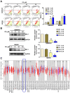
PD treatment induces mitochondrial dysfunction in human glioblastoma cells. GBM8401 and M059K cells were treated with various concentrations of PD for 24 hours, and (A) MitoPotential staining was performed to observe to MMPs. Right panel: quantification of MMPs. (B) Western blot analysis of MCL-1 expression. Right panel: quantification of fold-changes in MCL-1/GAPDH. (C) TCGA data analysis using Time2.0 software compared MCL-1 expression in normal and glioblastoma tissues. Data are presented as the mean ± SD. **p < 0.01 vs. control cells.
Figure 4
Figure 4
PD induces autophagy by targeting LC3 in human glioblastoma cells. (A) GBM8401 and M059K glioblastoma cells were treated with various concentrations of PD for 24 hours, and the formation of acidic vesicular organelles was assessed using acridine orange staining. Total cell numbers were determined by nuclear Hoechst 33342 staining. (B) Western blot analysis of LC3B protein expression in GBM8401 and M059K cells. GAPDH was used as a loading control. Right panel: quantification of fold-changes in LC3B/GAPDH. Cells were pre-treated with siLC3 (20 nM) for 6 hours, then treated with PD (18 μM) for 24 hours. (C) Changes in the number of apoptotic GBM8401 cells and (D) mitochondrial membrane potential was determined by flow cytometric analysis. (E) Western blot analysis of LC3B and cleaved PARP (c-PARP) in GBM8401 cells. Right panel: quantification of fold-changes in c-PARP/GAPDH. Data are presented as the mean ± SD. *p < 0.05; **p < 0.01 vs. control; #p < 0.05 vs. PD-treated cells. Scale bar=50 µm
Figure 5
Figure 5
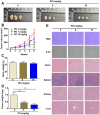
In vivo anti-tumor effect of PD in glioblastoma xenograft mice. (A) GBM8401 cells were subcutaneously injected into five-week-old female BALB/c nude mice, followed by oral administration of different concentrations of PD (5 and 10 mg/kg) for 5 weeks. Representative photographs of tumor-bearing mice are shown (n = 5 per group). (B) Tumor growth curves (tumor volume) under PD treatment were evaluated weekly for 5 weeks. (C) Body weight and (D) tumor weight were measured at the end of the experiment. (E) Tumor morphology was assessed by H&E staining, and Ki-67 expression was evaluated by immunohistochemical analysis. Drug safety was assessed by evaluating PD-induced organ toxicity (heart, spleen, kidney, and liver) using H&E staining. Data are presented as the mean ± SD. *p < 0.05; **p < 0.01 vs. control group. Scale bar=100 µm
Figure 6
Figure 6
PD directly targets LC3B/MCL-1 in molecular docking model. (A, C) The 3D structure of LC3B is shown with PD (yellow) docked into the binding site, indicating strong binding interaction. (D, F) Surface representation of LC3B and MCL-1 with PD bound to a hydrophobic pocket and shown in key interaction residues. (B, E) Docking scores of PD with LC3B and MCL-1 show the highest binding affinity. (G) Schematic diagram illustrates the proposed molecular mechanism of PD targeting LC3B and MCL-1 by which PD exerts anti-glioblastoma effects.

References

    1. Siminska D, Korbecki J, Kojder K, Kapczuk P, Fabianska M, Gutowska I. et al. Epidemiology of Anthropometric Factors in Glioblastoma Multiforme-Literature Review. Brain Sci. 2021;11:116. - PMC - PubMed
    1. Cruz-Martins N. Advances in Plants-Derived Bioactives for Cancer Treatment. Cells. 2023;12:1112. - PMC - PubMed
    1. Tagde P, Tagde P, Tagde S, Bhattacharya T, Garg V, Akter R. et al. Natural bioactive molecules: An alternative approach to the treatment and control of glioblastoma multiforme. Biomed Pharmacother. 2021;141:111928. - PubMed
    1. Navarro Del Hierro J, Casado-Hidalgo G, Reglero G, Martin D. The hydrolysis of saponin-rich extracts from fenugreek and quinoa improves their pancreatic lipase inhibitory activity and hypocholesterolemic effect. Food Chem. 2021;338:128113. - PubMed
    1. He X, Qiao A, Wang X, Liu B, Jiang M, Su L. et al. Structural identification of methyl protodioscin metabolites in rats' urine and their antiproliferative activities against human tumor cell lines. Steroids. 2006;71:828–33. - PubMed

MeSH terms