. 2025 Oct 3:8:0937.
doi: 10.34133/research.0937. eCollection 2025.

Noninvasive Computed Tomography-Based Quantification of Tumor Fibrosis Predicts Pancreatic Cancer Response to Gemcitabine/Nab-Paclitaxel

Affiliations

Noninvasive Computed Tomography-Based Quantification of Tumor Fibrosis Predicts Pancreatic Cancer Response to Gemcitabine/Nab-Paclitaxel

Qiuxia Yang et al. Research (Wash D C). .

Abstract

Pancreatic ductal adenocarcinoma (PDAC) carries a dismal prognosis. Chemotherapy remains the mainstay for unresectable cases, yet regimens like AG (gemcitabine/nab-paclitaxel) exhibit heterogeneous efficacy. Tumor fibrosis has emerged as a potential predictor of treatment response but lacks validated noninvasive assessment methods. To address this, in this multicenter study, tumor fibrosis was quantified in 361 patients with resectable PDAC from SYSUCC, XYCSU, and TCGA cohorts using deep learning-based tissue segmentation on hematoxylin and eosin-stained whole-slide images. Fibrosis was defined as stromal proportion, and its association with overall survival (OS) was evaluated. Transcriptomic profiling was performed in 51 XYCSU cases to validate the biological relevance of fibrosis quantification. A radiomics model (RM) was then developed using preoperative contrast-enhanced computed tomography (CT) scans from SYSUCC to predict fibrosis and externally validated in XYCSU. Clinical utility was assessed in an independent cohort of 295 unresectable PDAC patients treated with AG, FOLFIRINOX, or SOXIRI. High fibrosis correlated with prolonged OS across resectable cohorts (all P < 0.05). Transcriptomic analysis revealed enrichment of fibrosis-related pathways in high-fibrosis tumors. The RM achieved an area under the curve of 0.718 (95% confidence interval: 0.627 to 0.823) in the external test set. Among patients receiving AG, those with CT-predicted high fibrosis had significantly longer progression-free survival (median: 6.23 versus 4.70 months, P = 0.037) and OS (13.37 versus 7.73 months, P = 0.002). No significant survival benefit was observed for high-fibrosis patients receiving FOLFIRINOX or SOXIRI. CT-based fibrosis quantification offers a robust, noninvasive biomarker for predicting AG efficacy in unresectable PDAC.


Conflict of interest statement

Competing interests: The authors declare that they have no competing interests.

Figures
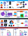
Fig. 1.
Fig. 1.
Overview of the study design.
Fig. 2.
Fig. 2.
Flowchart of patient screening and enrollment for each study cohort. Different cohorts are collected for the following tasks: (A) WSI-based fibrosis assessment and CT-based fibrosis prediction model, (B) clinical relevance of CT-predicted fibrosis across chemotherapy regimens, and (C) differentially expressed gene profiling analysis. PDAC, pancreatic ductal adenocarcinoma; H&E, hematoxylin-eosin; WSI, whole-slide image; AG, gemcitabine/nab-paclitaxel; FOLFIRINOX, 5-fluorouracil, leucovorin, irinotecan, and oxaliplatin; SOXIRI, S-1, oxaliplatin, and irinotecan; M1, metastatic pancreatic ductal adenocarcinoma.
Fig. 3.
Fig. 3.
WSI-based fibrosis assessment. (A) Schematic illustration of manual annotations for 8 distinct tissue classes in whole-slide image. (B) Development of tissue classification models and visualization of segmentation results. (C) Representative segmentation maps from high- and low-fibrosis patients, with stroma shown in green. (D) WSI-based fibrosis stratification is significantly associated with overall survival across all cohorts.
Fig. 4.
Fig. 4.
Fibrosis-associated differentially expressed gene profiling. (A) Analytical workflow. Fifty-one PDAC patients from XYCSU with transcriptomic data were stratified into high- and low-fibrosis groups based on WSI-derived fibrosis assessment. (B) Enrichment network visualization. Nodes represent significantly enriched gene sets, with node size proportional to gene count. Edges indicate overlap between gene sets, with thickness scaled to shared gene count. (C) Volcano plot of 364 fibrosis-associated DEGs (red: up-regulated in high-fibrosis; blue: down-regulated).
Fig. 5.
Fig. 5.
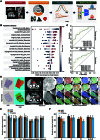
CT-based fibrosis prediction. (A) Workflow of radiomics analysis. (B) SHAP summary plot showing the top 15 radiomic features contributing to fibrosis prediction. (C) Fivefold cross-validation performance of the CT-based fibrosis prediction model on the training (SYSUCC) and test (XHCSU) cohorts. (D) Representative contrast-enhanced CT (CECT) images with discriminative textural feature maps in high- and low-fibrosis patients. (E) Performance comparison of different fibrosis prediction models using features extracted from venous-phase CT images. (F) Performance comparison of different CT imaging phases. SVM classifiers were built using features extracted from arterial-, venous-, and delayed-phase images, as well as their average combination. AUC, area under the receiver operating characteristic curve; ACC, accuracy; A, arterial phase image; V, venous phase image; D, delayed phase image.
Fig. 6.
Fig. 6.
Clinical utility of CT-based fibrosis prediction across chemotherapy regimens. (A) Overall survival stratified by CT-predicted fibrosis status (high versus low) in patients receiving AG, FOLFIRINOX, or SOXIRI. (B) Progression-free survival stratified by CT-predicted fibrosis status (high versus low) under the same treatment regimens.
Fig. 7.
Fig. 7.
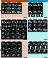
Serial CECT assessment of treatment response stratified by fibrosis status (high versus low) and chemotherapy regimens (AG, FOLFIRINOX, and SOXIRI). The detailed legends of each image were described in Supplementary Methods.

References

    1. Siegel RL, Kratzer TB, Giaquinto AN, Sung H, Jemal A. Cancer statistics, 2025. CA Cancer J Clin. 2025;75(1):10–45. - PMC - PubMed
    1. Lei S, Mao Y, Yang Q, Yan H, Wang J. Trends in pancreatic cancer incidence, prevalence, and survival outcomes by histological subtypes: A retrospective cohort study. Gastroenterol Rep. 2025;13: Article goaf030. - PMC - PubMed
    1. Gyawali B, Booth CM. Treatment of metastatic pancreatic cancer: 25 years of innovation with little progress for patients. Lancet Oncol. 2024;25(2):167–170. - PubMed
    1. Hu ZI, O’Reilly EM. Therapeutic developments in pancreatic cancer. Nat Rev Gastroenterol Hepatol. 2024;21(1):7–24. - PubMed
    1. Mastrantoni L, Chiaravalli M, Spring A, Beccia V, Di Bello A, Bagalà C, Bensi M, Barone D, Trovato G, Caira G, et al. Comparison of first-line chemotherapy regimens in unresectable locally advanced or metastatic pancreatic cancer: A systematic review and Bayesian network meta-analysis. Lancet Oncol. 2024;25(12):1655–1665. - PubMed

LinkOut - more resources