. 2025 Oct 7;10(1):333.
doi: 10.1038/s41392-025-02430-5.

Z-DNA-binding protein 1 exacerbates myocardial ischemia‒reperfusion injury by inducing noncanonical cardiomyocyte PANoptosis

Affiliations

Z-DNA-binding protein 1 exacerbates myocardial ischemia‒reperfusion injury by inducing noncanonical cardiomyocyte PANoptosis

Xiaokai Zhang et al. Signal Transduct Target Ther. .

Abstract

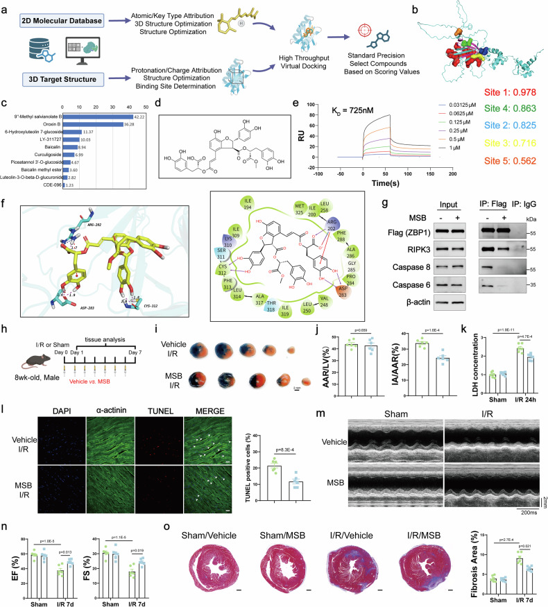
Myocardial ischemia‒reperfusion (I/R) injury is the primary factor that counteracts the beneficial effects of reperfusion therapy. Cardiomyocyte death serves as the fundamental pathological hallmark of I/R injury. However, targeting a single type of cell death has been reported to be ineffective at preventing I/R injury. ZBP1 is well established as a nucleic acid sensor that activates inflammatory and various cell death signaling pathways. However, the specific role of ZBP1 in adult cardiomyocytes, particularly in the absence of nucleic acid ligands, remains largely unexplored. In this study, our dynamic transcriptomic analyses at various I/R stages revealed a cluster of genes significantly enriched in cell death-related processes, with ZBP1 showing significant expression changes in both our I/R injury mouse model and public human ischemic cardiomyopathy datasets. Cardiomyocytes are the primary cell type expressing ZBP1 in response to I/R injury. Hypoxia/reoxygenation stress induced the upregulation of multiple cell death markers indicative of PANoptosis in adult cardiomyocytes, which was mitigated by ZBP1 deficiency. Compared with treatment with conventional cell death inhibitors, cardiomyocyte-specific Zbp1 deficiency ameliorated I/R-induced PANoptosis, resulting in a more substantial reduction in myocardial infarct size. Conversely, myocardial Zbp1 overexpression in adult mice directly induced cardiac remodeling and heart failure. Mechanistically, ZBP1 drives cardiomyocyte PANoptosis by promoting the formation of the ZBP1/RIPK3/CAS8/CAS6 PANoptosome complex. Virtual screening and experimental validation revealed a novel small-molecule compound, MSB, which has high binding affinity for ZBP1 and effectively attenuates myocardial I/R injury both in vitro and in vivo. Collectively, these findings highlight the role of ZBP1 as a mediator of cardiomyocyte PANoptosis and suggest that targeting ZBP1 could be a promising strategy for mitigating myocardial I/R injury.


Conflict of interest statement

Competing interests: The authors declare no competing interests.

Figures

Fig. 1
Fig. 1
Integrative bioinformatics analyses identified an essential role of ZBP1 in myocardial I/R injury. a Schematic diagram showing the mouse model of I/R injury. b Principal component analysis (PCA) showing group distribution on the basis of RNA-seq analyses of myocardial samples from the ischemic regions of wild-type (WT) mice subjected to sham surgery or after 30 min of ischemia followed by reperfusion (duration 0, 1, 6, and 24 h). c The dynamic change patterns of differentially expressed genes (DEGs) remained unchanged at 0 h post-I/R but were consistently upregulated at 0, 1, 6, and 24 h post-I/R. d Heatmap of DEGs that remained unchanged at 0 h post-I/R but were consistently upregulated at 0, 1, 6, and 24 h post-I/R. e Schematic illustration of the screening process for candidate genes. f Quantitative real-time polymerase chain reaction (qRT‒PCR) analyses of Zbp1 mRNA levels in heart samples from WT mice at 0, 1, 6, and 24 h post-I/R or sham surgery (n = 6). g Western blot analysis of ZBP1 protein levels normalized to that of β-actin (ZBP1/actin ratio) in heart samples from normal controls and ICM patients (n = 6 for normal controls and n = 8 for ICM patients). h Representative immunohistochemical staining and quantification of ZBP1 in heart samples from normal controls and ICM patients (n = 6 for normal controls and n = 8 for ICM patients; scale bar = 20 μm). The hematoxylin-stained nuclei appeared blue, while the DAB-positive signal was brown-yellow. i Quantitative real-time polymerase chain reaction (qRT‒PCR) analysis of Zbp1 mRNA levels in cardiomyocytes isolated from WT mice 24 h post-I/R or sham surgery (n = 6). j Western blot of ZBP1 in cardiomyocytes isolated from WT mice 24 h post-I/R or sham surgery (n = 3). k Immunofluorescence of 4′,6-diamidino-2-phenylindole (DAPI; blue), ZBP1 (green) and α-actinin (red) in the myocardium of WT mice 24 h post-I/R or sham surgery (scale bar = 20 μm in the left panel and 10 μm in the right panel). For all the statistical plots, the data are presented as the means ± SEMs. f by one-way ANOVA with Bonferroni multiple comparison test. g by two-tailed unpaired Student’s t-test. h, i by Welch’s t-test
Fig. 2
Fig. 2
Zbp1 knockdown attenuates pyroptosis, apoptosis, and necroptosis in cardiomyocytes after H/R stress. a Quantitative real-time polymerase chain reaction (qRT‒PCR) analyses of Zbp1 mRNA levels in adolescent cardiomyocytes subjected to H/R for different durations (n = 4). b Immunofluorescence of ZBP1 in adult Myh6+ and Zbp1fl/flMyh6+ mouse cardiomyocytes subjected to H/R for 0.5/24 h (scale bar = 20 μm). c Western blot and quantification of ZBP1, total caspase 3 (T-Caspase 3), cleaved caspase 3 (Cl-Caspase 3), phosphorylated-RIPK3 (p-RIPK3), RIPK3, full-length GSDMD (GSDMD-FL) and GSDMD-N fragment protein levels in adult Myh6+ and Zbp1fl/flMyh6+ mouse cardiomyocytes subjected to H/R for different durations (n = 4). d Microscopy images and quantification of adult cardiomyocytes induced by H/R for 0.5/24 h were obtained via propidium iodide (PI) and Hoechst staining (n = 4; scale bar = 50 μm). e LDH (lactate dehydrogenase) release by adult Myh6+ and Zbp1fl/flMyh6+ mouse cardiomyocytes subjected to H/R for 0.5–24 h (n = 4). f Caspase-3 activity in the cardiomyocytes of adult Myh6+ and Zbp1fl/flMyh6+ mice subjected to H/R for 0.5/24 h (n = 4). For all the statistical plots, the data are presented as the means ± SEMs. a by one-way ANOVA with Bonferroni multiple comparison test. c and e, f by two-way ANOVA with Bonferroni multiple comparison test. d by Welch’s ANOVA with Dunnett’s T3 multiple comparison test
Fig. 3
Fig. 3
Cardiomyocyte-specific Zbp1 deficiency ameliorates I/R-induced myocardial injury in vivo. a Schematic diagram showing the mouse model of I/R injury. The Myh6+ and Zbp1fl/flMyh6+ mice were intraperitoneally injected with Z-VAD-FMK (20 mg/kg), GSK’872 (10 mg/kg) or vehicle control the day before I/R surgery. b Representative images of heart sections from the indicated groups of mice stained with Evans blue and TTC at 24 h after myocardial-ischemia‒reperfusion injury. c The ratios of the area at risk (AAR) to the left ventricle (LV) and the infarct area (IA) to the AAR were calculated via Evans blue and TTC staining (n = 6). d The serum levels of lactate dehydrogenase (LDH, U/L) in the indicated groups of mice 24 h after I/R surgery (n = 8). e Representative images and quantitative analysis of myocardial Evans blue dye (EBD) uptake and viable cardiomyocytes labeled with α-actinin antibodies in Myh6+ and Zbp1fl/flMyh6+ mice 24 h after I/R surgery (n = 10; scale bar = 100 μm). f Representative TUNEL staining and relative quantification of heart samples from Myh6+ and Zbp1fl/flMyh6+ mice 24 h after I/R surgery (n = 10; scale bar = 20 μm). g Western blot and quantification of ZBP1, total caspase 3 (T-Caspase 3), cleaved caspase 3 (Cl-Caspase 3), phosphorylated-RIPK3 (p-RIPK3), RIPK3, full-length GSDMD (GSDMD-FL) and GSDMD-N fragment protein levels in heart samples from Myh6+ and Zbp1fl/flMyh6+ mice 24 h after I/R surgery (n = 4). For all the statistical plots, the data are presented as the means ± SEMs. c, d by one-way ANOVA with Bonferroni multiple comparison test. e, f by two-tailed unpaired Student’s t-test. g by two-way ANOVA with Bonferroni multiple comparison test
Fig. 4
Fig. 4
Cardiomyocyte-specific overexpression of Zbp1 leads to cardiac remodeling and heart failure. a The survival rates of Myh6+ and Zbp1TGMyh6+ mice were estimated via the Kaplan‒Meier method. b M-mode echocardiography of Myh6+ and Zbp1TGMyh6+ mice 8 weeks post-tamoxifen injection. c, d Left ventricular EF, FS (c) and LVIDs (d) were assessed by echocardiography in Myh6+ and Zbp1TGMyh6+ mice 8 weeks after tamoxifen injection (n = 10). e Ratios of HW to BW and LW to TL in Myh6+ and Zbp1TGMyh6+ mice 8 weeks after tamoxifen injection (n = 6). f Representative images and quantitative analysis of Masson staining of heart sections from Myh6+ and Zbp1TGMyh6+ mice 8 weeks post-tamoxifen injection (n = 10; scale bar = 500 µm in the upper panel and 50 µm in the lower panel). g Western blot and quantification of ZBP1, BNP, MYH7 and Collagen I protein levels in heart samples from Myh6+ and Zbp1TGMyh6+ mice (n = 6). h Representative photographs and quantitative analysis of myocardial Evans blue dye (EBD) uptake and WGA (wheat germ agglutinin) staining in Myh6+ and Zbp1TGMyh6+ mice 8 weeks post-tamoxifen injection (n = 10; scale bar = 20 μm). Representative TUNEL staining and relative quantification of heart samples from Myh6+ and Zbp1TGMyh6+ mice 8 weeks post-tamoxifen injection (n = 10; scale bar = 20 μm). i Western blot and quantification of phosphorylated-RIPK3 (p-RIPK3) and RIPK3 protein levels in cardiomyocytes isolated from Myh6+ and Zbp1TGMyh6+ mice 8 weeks after tamoxifen injection (n = 6). j Western blot and quantification of total caspase 3 (T-Caspase 3) and cleaved caspase 3 (Cl-Caspase 3) protein levels in cardiomyocytes isolated from Myh6+ and Zbp1TGMyh6+ mice 8 weeks after tamoxifen injection (n = 6). k Western blot and quantification of full-length GSDMD (GSDMD-FL) and GSDMD-N fragment protein levels in cardiomyocytes isolated from Myh6+ and Zbp1TGMyh6+ mice 8 weeks after tamoxifen injection (n = 6). l Western blot and quantification of total caspase 8 (T-Caspase 8) and cleaved caspase 8 (Cl-Caspase 8) protein levels in cardiomyocytes isolated from Myh6+ and Zbp1TGMyh6+ mice 8 weeks after tamoxifen injection (n = 6). m Western blot and quantification of phosphorylated RIPK1 (p-RIPK1) and RIPK1 protein levels in cardiomyocytes isolated from Myh6+ and Zbp1TGMyh6+ mice 8 weeks post-tamoxifen injection (n = 6). For all the statistical plots, the data are presented as the means ± SEMs. a by the log-rank test. ce and im by two-tailed unpaired Student’s t-test. fh by Welch’s t-test. EF ejection fraction, FS fractional shortening, LVIDs left ventricular internal diameter at end-systole, HW/BW ratio of heart weight to body weight, LW/TL ratio of lung weight to tibia length
Fig. 5
Fig. 5
ZBP1 Induces cardiomyocyte PANoptosis by modulating noncanonical PANoptosome assembly. a Coimmunoprecipitation and Western blot assays of Flag (ZBP1) and RIPK3, Caspase 8, Caspase 6, cGAS, RIPK1, NLRP3, and ASC interaction in cardiomyocytes infected with adenovirus expressing ZBP1. b Coimmunoprecipitation and Western blot assays of Flag (ZBP1) and RIPK3, Caspase 8, and Caspase 6 in cardiomyocytes isolated from WT or cGAS-/- mice infected with adenovirus expressing ZBP1. c Coimmunoprecipitation and Western blot assays of RIPK3 and ZBP1, Caspase 8, and Caspase 6 interaction in cardiomyocytes infected with adenovirus expressing ZBP1. d Coimmunoprecipitation and Western blot assays of the interaction between Caspase 6 and ZBP1, RIPK3, and Caspase 8 in cardiomyocytes infected with adenovirus expressing ZBP1. e Representative confocal images of ZBP1 and RIPK3, Caspase 8, Caspase 6 colocalization in heart sections from Myh6+ and Zbp1TGMyh6+ mice 8 weeks after tamoxifen injection (scale bar = 10 μm). f Coimmunoprecipitation and Western blot analyses of RIPK3 and RIPK1, Caspase 8, and Caspase 6 interactions in adult Myh6+ and Zbp1fl/flMyh6+ mouse cardiomyocytes subjected to H/R for 0.5/24 h. g Coimmunoprecipitation and Western blot assays of Flag (ZBP1) and RIPK3, Caspase 6, and Caspase 8 interaction in cardiomyocytes infected with adenovirus expressing sh-RIPK3, sh-Caspase 6 or sh-Caspase 8 with ZBP1 overexpression. h Western blot of ZBP1, total caspase 6 (T-Caspase 6) and cleaved caspase 6 (Cl-Caspase 6) in cardiomyocytes infected with adenovirus expressing ZBP1 or the NC
Fig. 6
Fig. 6
MSB is a novel ZBP1 inhibitor that protects against myocardial ischemia‒reperfusion injury. a Schematic diagram displaying a strategy to screen mouse ZBP1 inhibitors. b The druggable binding pockets of mouse ZBP1 (AlphaFold ID: AF-Q9QY24-F1) were predicted and scored on the basis of its 3D structure. Virtual screening was performed according to site 4. c Surface plasmon resonance (SPR) analysis of the binding response of ten compounds with ZBP1 at a single concentration (50 µM). d The chemical structure of MSB. e Kinetic curves from subsequent concentration-dependent SPR analysis showing the binding of MSB to ZBP1. f 2D and 3D patterns of MSB and mouse ZBP1 binding. g Coimmunoprecipitation and Western blot assays of Flag (ZBP1) and RIPK3, Caspase 6, and Caspase 8 interactions in cardiomyocytes infected with adenovirus expressing ZBP1 with or without MSB (1 μM) treatment. h Schematic representation of the experimental protocol for myocardial I/R injury in mice treated with vehicle or MSB (5 mg/kg/day, 2 h before the onset of ischemia for the first time). i Representative images of heart sections stained with Evans blue and TTC from WT mice 24 h after I/R surgery and treated with MSB or vehicle. j The ratios of the area at risk (AAR) to the left ventricle (LV) and the infarct area (IA) to the AAR were calculated via Evans blue and TTC staining (n = 6). k Serum levels of lactate dehydrogenase (LDH, U/L) in WT mice 24 h after I/R surgery treated with MSB or vehicle (n = 6). l Representative TUNEL staining and relative quantification of heart samples from WT mice 24 h after I/R surgery and treated with MSB or vehicle (n = 6; scale bar = 20 μm). m M-mode echocardiography of WT mice 7 days post-I/R injury treated with MSB or vehicle. n Left ventricular EF and FS were assessed by echocardiography in WT mice 7 days post-I/R injury treated with MSB or vehicle (n = 6). o Representative images and quantitative analysis of Masson staining of heart sections from WT mice 7 days post-I/R injury treated with MSB or vehicle (n = 6, scale bar = 500 μm). For all the statistical plots, the data are presented as the means ± SEMs. j and l by two-tailed unpaired Student’s t-test. k and no by two-way ANOVA with Bonferroni multiple comparison test. EF ejection fraction, FS fractional shortening
Fig. 7
Fig. 7
A schematic illustration showing that ZBP1 drives cardiomyocyte PANoptosis through the formation of the ZBP1/RIPK3/CASP6/CASP8 complex. Myocardial I/R injury induced the upregulation of ZBP1 in adult cardiomyocytes. ZBP1 facilitates the assembly of the ZBP1/RIPK3/CAS8/CAS6 PANoptosome complex, thereby driving multiple forms of programmed cell death, including pyroptosis, apoptosis, and necroptosis. MSB exhibits high binding affinity for ZBP1 and effectively attenuates myocardial I/R injury. The schematic illustration was generated using BioRender (https://BioRender.com)

References

    1. Anderson, J. L. & Morrow, D. A. Acute myocardial infarction. N. Engl. J. Med.376, 2053–2064 (2017). - PubMed
    1. Heusch, G. Myocardial ischemia/reperfusion: translational pathophysiology of ischemic heart disease. Med.5, 10–31 (2024). - PubMed
    1. Heusch, G. Myocardial ischaemia-reperfusion injury and cardioprotection in perspective. Nat. Rev. Cardiol.17, 773–789 (2020). - PubMed
    1. Xiang, Q., Yi, X., Zhu, X. H., Wei, X. & Jiang, D. S. Regulated cell death in myocardial ischemia-reperfusion injury. Trends Endocrinol. Metab.35, 219–234 (2024). - PubMed
    1. Del Re, D. P., Amgalan, D., Linkermann, A., Liu, Q. & Kitsis, R. N. Fundamental mechanisms of regulated cell death and implications for heart disease. Physiol. Rev.99, 1765–1817 (2019). - PMC - PubMed

LinkOut - more resources