. 2025 Oct 7;8(12):e202503414.
doi: 10.26508/lsa.202503414. Print 2025 Dec.

Beneficial and detrimental consequences of AHR activation in intestinal infection

Affiliations

Beneficial and detrimental consequences of AHR activation in intestinal infection

Oscar E Diaz et al. Life Sci Alliance. .

Abstract

The ligand-dependent transcription factor aryl hydrocarbon receptor (AHR) is an environmental sensor whose activation can have physiologically beneficial or detrimental consequences for host immune responses depending on the ligand. Here, we investigated the hypothesis that prolonged AHR activation either because of inefficient ligand metabolism or because of genetic manipulation may underlie the distinction between beneficial and detrimental effects. Our data indicate that prolonged AHR activation caused toxic endpoints for liver and thymus but was not per se interfering with the host response to infection with the intestinal pathogen C. rodentium Genetically driven constitutive AHR activation improved resistance to infection, whereas prolonged AHR activation by the pollutant TCDD resulted in delayed clearance of C. rodentium associated with a suppression in antibody production. Combined single-cell RNA-seq and ATAC-seq analysis provided evidence that TCDD, but not genetic AHR activation, negatively affected dendritic cell functions such as activation, maturation, and antigen presentation. Thus, the detrimental impact of environmental pollutants such as TCDD on immune responses cannot solely be attributed to aberrantly prolonged activation of AHR.


Conflict of interest statement

The authors declare that they have no conflict of interest.

Figures

Figure 1.
Figure 1.. AHR expression in intestinal immune cell populations.
(A) AHR-TdTomato expression across immune cell types in the colon lamina propria and colon-draining mesenteric lymph node was determined by flow cytometry. Each dot represents the geometric MFI in the respective cell type. (B) Percentage of AHR-TdTomato–positive cells in each immune cell type shown in (A). Each dot represents the frequency of AHR-TdTomato+ cells in the respective cell type, n = 2 mice per group, and the bars show the mean + SD. Data are representative of three experiments.
Figure S1.
Figure S1.. AHR expression in intestinal immune cell populations.
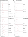
(A, B) Histograms showing AHR-TdTomato expression of immune cell types in the colon lamina propria and c-MLN in Fig 1 determined by flow cytometry. Histograms show concatenated events from cells obtained from n = 2 mice per group. Grey = WT control, Red = AHR-TdTomato.
Figure S2.
Figure S2.. (A, B) Gating strategy for immune cell populations from the (A) colon and (B) c-MLN in Ahr-TdTomato mice.
Numbers show percentage of cells gated from all events in the respective dotplot. A plot on the right shows the gating for defining AHR-TdTomato+ cells in a representative cell type.
Figure 2.
Figure 2.. Prolonged AHR activation.
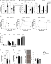
(A, B, C) Cyp1a1 and Ahrr gene expression was determined by qRT-PCR from the indicated intestinal sections from AhrdCAIR/dCAIR mice and WT mice (A) and 6 d after a single administration of 10 μg/kg TCDD (B, C), and presented as fold change relative to Hprt. Each dot represents one mouse, and the bars show the mean + SD. (A) WT, n = 10 males; AhrdCAIR/dCAIR = 12 males; 3 experiments. (B) Vehicle, n = 7 males; TCDD = 8 males; 2 experiments. (C) Vehicle, n = 3 males; TCDD, n = 3 males; 1 experiment. Data in (C) are representative of two experiments. (D) Cyp1a1 gene expression from the indicated organs was determined by qRT-PCR. Gonadal white adipose tissue = gWAT. Timepoints correspond to the hours after a single administration of 250 mg/kg I3C or 10 μg/kg TCDD in corn oil. Expression values were normalized to Hprt and to values from the vehicle-treated mice. Each dot represents the mean of 2–3 mice + SD. (E) TCDD levels detected in the indicated organ and timepoint after a single administration of 10 μg/kg TCDD in corn oil normalized to the organ weight. Each dot represents one mouse for adipose and liver samples (n = 3 per group) and a pool of samples from three mice for the serum, small intestine (duodenum and ileum), and colon. (F, G) Weight of thymus and liver from AhrdCAIR/dCAIR or TCDD-treated mice relative to body weight. Each dot represents one mouse, and the bars show the mean + SD. (H) Representative Oil Red O (ORO) staining in liver sections of AhrdCAIR/dCAIR mice and TCDD-treated mice. Scale bars, 50 μm (left panel). Quantification of ORO stain (right panel). Each dot represents one mouse, and the bars show the mean + SD. (F, G, H) WT, n = 8 females; TCDD, n = 6 females; AhrdCAIR/dCAIR, n = 3 females; 2 experiments. *P < 0.05, **P < 0.01, ***P < 0.001, ****P < 0.0001. Unpaired t test (A, B), Two-way ANOVA with Šídák’s multiple comparisons test. (C, D), One-way ANOVA with Tukey’s multiple comparisons test (F, G, H).
Figure 3.
Figure 3.. Impact of prolonged AHR activation on Citrobacter rodentium infection.
(A, B, C) C. rodentium burden in faecal pellets, (B) IL-22 protein levels in supernatants from colon explant cultures, (C) Lcn2 protein levels in faecal pellets from AhrdCAIR/dCAIR and WT mice. (A, B, C) WT, n = 16 males; AhrdCAIR/dCAIR, n = 13 males; 5 experiments; (B) WT, n = 10 males; AhrdCAIR/dCAIR, n = 3 males; 2 experiments; and (C) WT, n = 12 males; AhrdCAIR/dCAIR, n = 5 males; 3 experiments. (D, E, F) C. rodentium burden in faecal pellets, (E) IL-22 protein levels in supernatants from colon explant cultures and (F) Lcn2 protein levels in faecal pellets from TCDD-treated and vehicle-treated mice. Mice were infected 6 d after TCDD or vehicle administration. (D, E, F) Vehicle, n = 4 males; TCDD, n = 4 males; 1 experiment and representative of at least two experiments. Each dot represents samples collected from one mouse, and the bars show the mean + SD. Two-way ANOVA with Šídák’s multiple comparisons test (A, C, D, F), Unpaired t test (B, E). *P < 0.05, **P < 0.01.
Figure S3.
Figure S3.. Impact of prolonged AHR activation to Citrobacter rodentium infection.
(A) C. rodentium burden in faecal pellets in female AhrdCAIR/dCAIR mice and WT mice (WT, n = 8 females; AhrdCAIR/dCAIR, n = 10 females, 3 experiments). (B) C. rodentium burden in faecal pellets in vehicle and TCDD-treated mice (vehicle, n = 6 females; TCDD, n = 4 females, 1 experiment). Each dot represents samples collected from one mouse and the bars show the mean + SD. (A, B) Two-way ANOVA with Šídák’s multiple comparisons test (A, B). *P < 0.05, **P < 0.01, ***P < 0.001.
Figure S4.
Figure S4.. Delayed clearance of Citrobacter rodentium in mice exposed to TCDD.
C. rodentium burden in faecal pellets of male mice infected with C. rodentium 6 d after an oral dose of 10 µg/kg TCDD or vehicle before infection. Vehicle, n = 4 males; TCDD, n = 5 males; 1 experiment. Data are representative of two experiments. Each dot corresponds to one mouse and bars represent the mean + SD. Two-way ANOVA with Šídák’s multiple comparisons test. *P < 0.05, **P < 0.01.
Figure 4.
Figure 4.. TCDD reduces levels of C. rodentium-specific antibodies.
(A) Levels of C. rodentium-specific Ig subclasses at the indicated serum dilutions 14 d after C. rodentium infection in mice that received 10 μg/kg TCDD or vehicle 6 d before infection. Vehicle, n = 6 females and 5 males; TCDD, n = 4 females and 6 males; 1 experiment. Data are representative of two experiments. Each dot represents samples collected from one mouse, and the bars show the mean + SD. (B) Levels of indicated Ig subclasses at the indicated serum dilutions 14 d after C. rodentium infection in AhrdCAIR/dCAIR and WT mice. WT, n = 3 females and 2 males; AhrdCAIR/dCAIR, n = 2 females and 2 males; 1 experiment. Data are representative of two experiments. Each dot represents one mouse, and the bars show the mean + SD. (A, B) Two-way ANOVA with Šídák’s multiple comparisons test (A, B). *P < 0.05.
Figure S5.
Figure S5.. TCDD suppresses levels of C. rodentium-specific antibodies.
(A) Levels of C.rodentium-specific Ig subclasses at the indicated sera dilutions 14 d after C. rodentium infection in mice that received 10 μg/kg TCDD or vehicle 6 d before infection. Vehicle, n = 6 females and 5 males; TCDD, n = 4 females and 6 males; 1 experiment. Data are representative of two experiments. Each dot represents samples collected from one mouse and the bars show the mean + SD. (B) Levels of indicated Ig subclasses at the indicated sera dilutions 14 d after C. rodentium infection in AhrdCAIR/dCAIR and WT mice. WT, n = 3 females and 2 males; AhrdCAIR/dCAIR, n = 2 females and 2 males; 1 experiment. Data are representative of two experiments. Each dot represents samples collected from one mouse and the bars show the mean + SD. (A, B) Two-way ANOVA with Šídák’s multiple comparisons test (A, B).
Figure S6.
Figure S6.. TCDD suppresses production of NP-specific IgG antibodies.
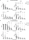
(A, B) Levels of NP-Ficoll and NP-CGG-specific IgG subclasses at the indicated serum dilutions 14 d after immunization with 10 μg NP-Ficoll or NP-CGG, respectively. Mice received 10 μg/kg TCDD or vehicle 6 d before immunization. Data are representative of two experiments (vehicle, n = 5 females and 5 males; TCDD, n = 5 females and 5 males; naïve = 1 female, 1 male). Each dot corresponds to one mouse, and the bars represent the mean + SD. (A, B) Two-way ANOVA with Šídák’s multiple comparisons test (A, B). *P < 0.05, **P < 0.01, ***P < 0.001, ****P < 0.0001.
Figure S7.
Figure S7.. TCDD affects immune cell composition in the c-MLN.
Cell numbers of total immune cells, and dendritic cell and B-cell populations in the c-MLN of mice 14 d after infection with C. rodentium 6 d after receiving 10 μg/kg TCDD or vehicle. Data were analysed by flow cytometry and represent one experiment (vehicle, n = 6 females; TCDD, n = 4 females). Each dot represents one mouse, and the bars show the mean + SD. t test. *P < 0.05, **P < 0.01, ***P < 0.001.
Figure 5.
Figure 5.. Single-cell RNA-seq analysis.
(A) Experimental setup for scMultiome from dendritic cells and CD45+ cells. (B) Dot plot showing the putative marker genes across cell types defined in the vehicle sample in the colon. The size of the dot represents the percentage of cells within clusters expressing the gene, whereas the colour scale represents the average expression level in that cluster. (C) Gene ontology enrichment analysis of biological processes for differentially expressed genes in TCDD and AhrdCAIR/dCAIR mice compared with vehicle (all males) for the indicated cell type. The colour scale represents the expression change bias, which represents the proportion of up-regulated and down-regulated genes that belong to that pathway, whereas the size of the dot shows the Z-score. Differentially expressed genes were defined as those with an adjusted P-value < 0.01, and enriched pathways have a P-value < 0.01. Abbreviations: Pep, peptide; Ag, antigen; proc, processing; pres, presentation; endog, endogenous; exog, exogenous; reg, regulation; pos, positive; transl, translation; init, initiation; stim, stimulatory; signal, signalling; imm, immune; resp, response; CAIR, AhrdCAIR/dCAIR.
Figure S8.
Figure S8.. Cell types identified in the single-cell RNA-seq and ATAC-seq datasets.
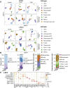
(A) Uniform Manifold Approximation and Projection using scRNA-seq or chromatin accessibility in vehicle, TCDD, and AhrdCAIR/dCAIR mice (referred to as CAIR in the figure). Cell types were assigned using the RNA-seq assay and visualized on both RNA- and ATAC-seq projections. (B) Proportion of dendritic cell subpopulations and of all other immune cells. (C) Dot plots adapted from Seurat DotPlot showing the putative marker genes across cell types defined in the vehicle sample in the c-MLN. The size of the dot represents the percentage of cells within a cell type expressing the marker gene, whereas colour represents the average scaled expression level of the marker gene in the cell type.
Figure S9.
Figure S9.. Gene ontology analysis of cDC2 populations in colon and c-MLN.
Gene ontology enrichment analysis of biological processes for differentially expressed genes in TCDD and AhrdCAIR/dCAIR mice compared with vehicle for the indicated cell type. The colour scale represents the expression change bias, which represents the proportion of up-regulated and down-regulated genes that belong to that pathway, whereas the size of the dot shows the Z-score. Abbreviations: Pep, peptide; Ag, antigen; proc, processing; pres, presentation; exog, exogenous; Pol, polymerase; reg, regulation; pos, positive; transl, translation; init, initiation; stim, stimulatory; signal, signalling; neg, negative; CAIR, AhrdCAIR/dCAIR.
Figure S10.
Figure S10.. Gene ontology analysis of cDC populations in colon and c-MLN.
Gene ontology enrichment analysis of biological processes for differentially expressed genes in AhrdCAIR/dCAIR mice compared with TCDD for the indicated cell type. The colour scale represents the expression change bias, which represents the proportion of up-regulated and down-regulated genes that belong to that pathway, whereas the size of the dot shows the Z-score. Abbreviations: Pep, peptide; Ag, antigen; proc, processing; pres, presentation; exog, exogenous; reg, regulation; pos, positive; transl, translation; init, initiation; stim, stimulatory; signal, signalling; neg, negative; imm, immune; resp, response; rec, receptor; CAIR, AhrdCAIR/dCAIR.
Figure 6.
Figure 6.. Gene expression and chromatin accessibility in cDC1 of AhrdCAIR/dCAIR mice compared with TCDD.
ATAC-seq signal profiles of differentially accessible peaks and linked genes that are also differentially expressed in cDC1 populations in colon and MLN. Differentially accessible peaks are shaded in grey, whereas violin plots show gene expression in the respective population and condition. ATAC-seq profiles were generated from pseudo-bulk chromatin accessibility data, and violin plots show normalized expression values at the single-cell level.

Update of

References

    1. Ahlfors H, Morrison PJ, Duarte JH, Li Y, Biro J, Tolaini M, Di Meglio P, Potocnik AJ, Stockinger B (2014) IL-22 fate reporter reveals origin and control of IL-22 production in homeostasis and infection. J Immunol 193: 4602–4613. 10.4049/jimmunol.1401244 - DOI - PMC - PubMed
    1. Akkaya M, Akkaya B, Sheehan PW, Miozzo P, Pena M, Qi CF, Manzella-Lapeira J, Bolland S, Pierce SK (2017) T cell-dependent antigen adjuvanted with DOTAP-CpG-B but not DOTAP-CpG-A induces robust germinal center responses and high affinity antibodies in mice. Eur J Immunol 47: 1890–1899. 10.1002/eji.201747113 - DOI - PMC - PubMed
    1. Andersson P, McGuire J, Rubio C, Gradin K, Whitelaw ML, Pettersson S, Hanberg A, Poellinger L (2002) A constitutively active dioxin/aryl hydrocarbon receptor induces stomach tumors. Proc Natl Acad Sci U S A 99: 9990–9995. 10.1073/pnas.152706299 - DOI - PMC - PubMed
    1. Anderton MJ, Manson MM, Verschoyle RD, Gescher A, Lamb JH, Farmer PB, Steward WP, Williams ML (2004) Pharmacokinetics and tissue disposition of indole-3-carbinol and its acid condensation products after oral administration to mice. Clin Cancer Res 10: 5233–5241. 10.1158/1078-0432.CCR-04-0163 - DOI - PubMed
    1. Bankoti J, Burnett A, Navarro S, Miller AK, Rase B, Shepherd DM (2010) Effects of TCDD on the fate of naive dendritic cells. Toxicol Sci 115: 422–434. 10.1093/toxsci/kfq063 - DOI - PMC - PubMed

MeSH terms

Substances

LinkOut - more resources