. 2025 Oct 7;11(1):81.
doi: 10.1038/s41421-025-00829-6.

Ferroptosis-induced SUMO2 lactylation counteracts ferroptosis by enhancing ACSL4 degradation in lung adenocarcinoma

Affiliations

Ferroptosis-induced SUMO2 lactylation counteracts ferroptosis by enhancing ACSL4 degradation in lung adenocarcinoma

Guangyao Shan et al. Cell Discov. .

Abstract

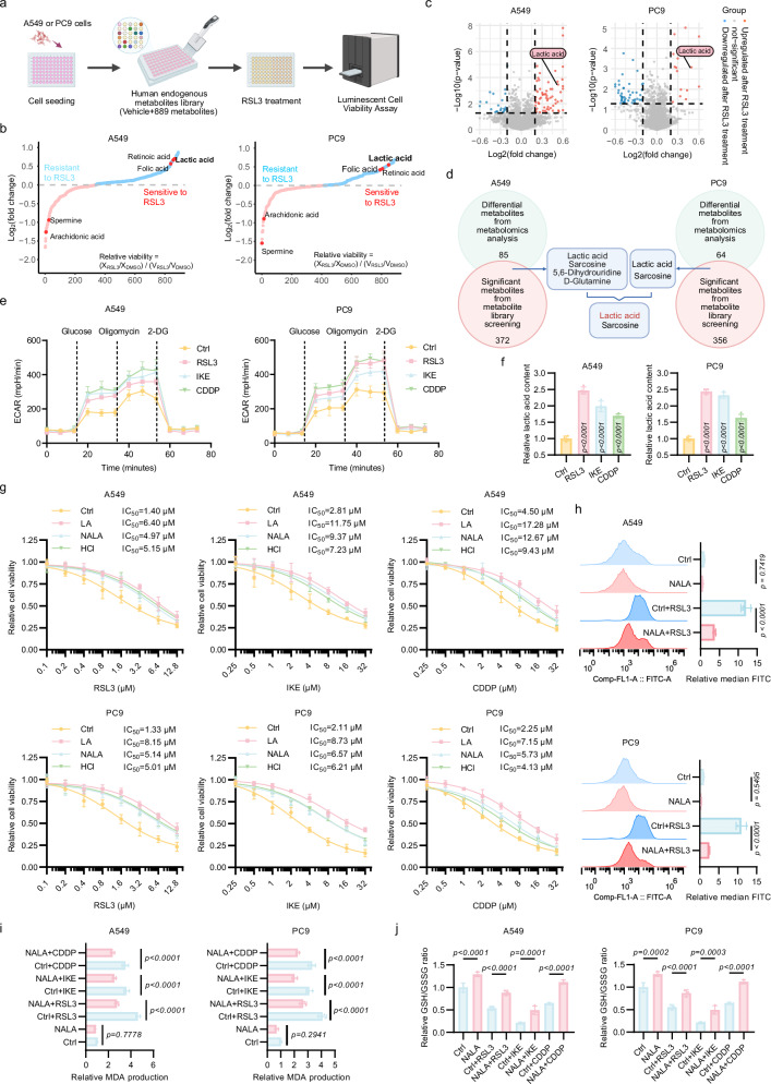
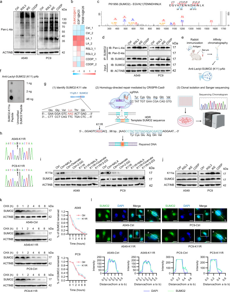
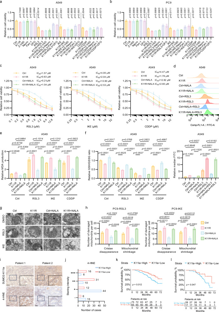
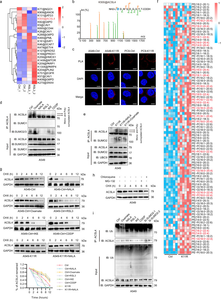
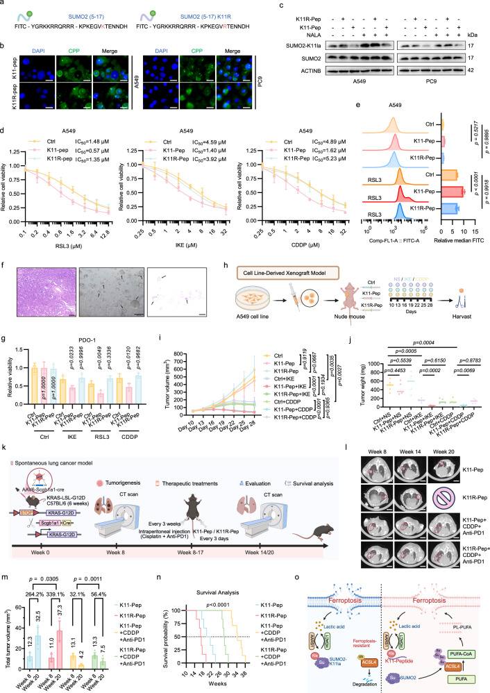
Lactylation, a lactate-mediated post-translational modification, has garnered significant attention for its pivotal role in epigenetic modulation. However, the intricate interplay between lactylation and ferroptosis in lung adenocarcinoma (LUAD) remains to be fully elucidated. Utilizing metabolomic profiling and comprehensive metabolic library screening, our study uncovers that ferroptosis markedly enhances lactic acid accumulation and subsequent protein lactylation, which in turn confers resistance to ferroptosis in LUAD cells. Functional assays, comprising cell viability tests, lipid peroxidation detection, as well as malondialdehyde and glutathione measurements, collectively reveal that SUMO2-K11 lactylation (SUMO2-K11la), the most prominently elevated lactylation in response to ferroptosis induction, serves as a pivotal factor in determining ferroptosis resistance. Sumoylation proteomics and co-immunoprecipitation assays reveal that SUMO2-K11la impairs the interaction between SUMO2 and ACSL4. Consequently, this disruption facilitates the degradation of ACSL4, thereby disrupting lipid metabolism and effectively mitigating ferroptosis. Furthermore, AARS1 is identified as the lactyltransferase and HDAC1 as the delactylase for SUMO2-K11la. Based on these findings, we develop a cell-penetrating peptide that competitively and specifically inhibits SUMO2-K11la. This peptide significantly potentiates ferroptosis and sensitizes LUAD to cisplatin in xenograft models, while enhancing chemoimmunotherapy responses in spontaneous lung cancer models. Overall, our findings imply that SUMO2-K11la is a pivotal regulator of ferroptosis resistance in LUAD, and suggest a promising strategy to potentiate ferroptosis-based cancer therapies via targeting SUMO2-K11la by the cell-penetrating peptide.


Conflict of interest statement

Competing interests: The authors declare no competing interests. Ethical approval: All participants have signed the informed consent. This study adhered to the Declaration of Helsinki and was approved by the Zhongshan Hospital Research Ethics Committee (B2022-180R).

Figures

Fig. 1
Fig. 1. Ferroptosis-induced lactic acid production contributes to ferroptosis resistance.
a Schematic workflow of human endogenous metabolite library screening in A549 and PC9 cells. b Relative viability of lung adenocarcinoma (LUAD) cells pre-treated with indicated metabolites (X) or vehicle control (V), followed by exposure to RSL3 (2 μM) or DMSO for 48 h. c Volcano plot of metabolomic profiling in A549 and PC9 cells post-RSL3 treatment (2 μM, 24 h). Metabolites with |log2(fold change)| > 0.2 and –log10(p-value) < 1.301 (dashed lines) were considered statistically significant. d Venn diagram highlighting overlapping metabolites identified by dual screening strategies as key regulators of ferroptosis. e Extracellular acidification rate (ECAR) in LUAD cells treated with RSL3 (2 μM), IKE (10 μM), or cisplatin (CDDP, 10 μM) for 24 h, measured via Seahorse XF Analyzer. f Lactic acid (LA) levels in LUAD cells following 24 h treatment with RSL3 (2 μM), IKE (10 μM), or CDDP (10 μM), quantified using an LA assay kit. g Dose-dependent effects of RSL3 (48 h), IKE (72 h), or CDDP (72 h) on A549 cell viability, with or without LA/Sodium lactate (NALA)/HCl supplementation (5 mM) (n = 4 biological replicates). hj Quantification of ferroptosis-associated biomarkers demonstrated the anti-ferroptotic activity of NALA: lipid-reactive oxygen species (lipid-ROS) were measured via BODIPY-C11 fluorescence probes (h), malondialdehyde (MDA) levels were assessed by thiobarbituric acid assay (i), and cellular redox status was determined by GSH/GSSG ratio (j). Data were analyzed by one-way ANOVA and were presented by mean ± SD.
Fig. 2
Fig. 2. Ferroptosis induces the lactylation of SUMO2 at lysine (K) 11 site.
a Pan-L-lactylation (Pan-L-Kla) levels were elevated in LUAD cells treated with RSL3 (2 μM), IKE (10 μM), or CDDP (10 μM) for 24 h. b Heatmap of three most significant lactylated protein sites identified by mass spectrometry (MS) in A549 cells treated with LA (20 mM), RSL3 (2 μM), or CDDP (10 μM), followed by immunoprecipitation with Pan-L-Kla antibody or IgG. c The B-y ion matching diagram of the lactylated SUMO2-K11 site (SUMO2-K11la). d Co-immunoprecipitation (Co-IP) assays showing enhanced SUMO2 lactylation level upon RSL3 (2 μM), IKE (10 μM), or CDDP (10 μM) treatment for 24 h. e Schematic diagram of customized SUMO2-K11la antibody development. f Dot blot validating the specificity for SUMO2-K11la antibody. g CRISPR-Cas9/homology-directed repair (HDR)-mediated generation of lactylation-deficient (Lysine 11 to Arginine, K11R) LUAD cells. h Sanger sequencing confirming the introduction of K11R mutations in A549 and PC9 cells. i SUMO2-K11la levels modulated by NALA (5 mM), sodium oxamate (5 mM), or 2-DG (5 mM) in Control (Ctrl) and K11R cells. j The SUMO2-K11la level was raised upon RSL3 (2 μM), IKE (10 μM), or CDDP (10 μM) treatment for 24 h. k Cycloheximide (CHX) chase assays showing unaltered SUMO2 protein stability in K11R mutants. l Immunofluorescence of SUMO2 (green) and DAPI (blue) confirming unchanged subcellular localization in K11R mutants. Scale bars, 20 μm. Data were presented by mean ± SD.
Fig. 3
Fig. 3. SUMO2-K11la counteracts ferroptosis in LUAD.
a, b Ferroptosis inhibitors ferrostatin-1 (Ferr-1, 10 μM) and deferoxamine (DFO, 10 μM) reversed K11R-driven cell death in LUAD cells, while apoptosis inhibitor Z-VAD-FMK (10 μM) and necroptosis inhibitor Necrosulfonamide (Necro, 10 μM) had no significant effect. c Relative viability of A549 Ctrl and K11R cells following treatment with varying concentrations of RSL3 (48 h), IKE (72 h), or CDDP (72 h) with or without concurrent incubation with NALA (5 mM). df Quantification of ferroptosis-associated biomarkers demonstrated the ferroptosis-promoting effect of K11R mutation: lipid-ROS were measured via BODIPY-C11 fluorescence probes (d), MDA levels were assessed by thiobarbituric acid assay (e), and cellular redox status was determined by GSH/GSSG ratio (f). g, h Transmission electron microscopy revealed pronounced mitochondrial damage (cristae disappearance and mitochondrial shrinkage) in K11R cells, which is not significantly affected by NALA supplementation. Scale bars, 5 μm. i, j Immunohistochemistry of LUAD patient tissues demonstrated an inverse correlation between SUMO2-K11la levels and lipid peroxidation marker 4-hydroxynonenal (4-HNE). Scale bars, 200 μm. k, l High SUMO2-K11la expression predicted reduced overall survival in 140 LUAD patients (k) and 45 CDDP-treated patients (l). Statistical analysis was performed using one-way ANOVA, and results were presented as mean ± SD.
Fig. 4
Fig. 4. SUMO2-K11la hinders the sumoylation of ACSL4 at K500 site to promote ACSL4 degradation.
a Quantitative SUMOylome profiling in LUAD cells under RSL3-induced ferroptosis (2 μM, 24 h) identified ACSL4-K500 as a dynamically regulated sumoylation site. b The B-y ion matching diagram of ACSL4-K500 sumoylated site. c Proximity ligation assay (PLA) confirmed enhanced SUMO2-ACSL4 interaction in SUMO2-K11R mutants. PLA: red; DAPI: blue. Scale bars, 10 μm. d Co-IP assays demonstrated suppression of SUMO2/3-mediated ACSL4 sumoylation by NALA or FINs, and its augmentation by sodium oxamate. e SUMO2-K11R mutation abolished lactylation-dependent regulation and hyper-sumoylated ACSL4. f Lipidomics revealed elevated PE-arachidonic acid (PE-AdA, 20:4) and PE-adrenic acid (PE-AA, 22:4) in SUMO2-K11R cells. g CHX chase assays showed accelerated ACSL4 degradation upon NALA/FIN treatment, reversed by sodium oxamate or SUMO2-K11R in A549 cells. h Proteasomal inhibitor MG132 (10 μM), but not lysosomal inhibitor chloroquine (20 μM), rescued ACSL4 degradation in A549 cells. i Ubiquitination assays indicated increased polyubiquitination of ACSL4 following NALA/FIN treatment or SUMO2 knockdown, contrasting with sodium oxamate-mediated suppression in A549 cells.
Fig. 5
Fig. 5. ACSL4-K500R mutation abrogates the effect of SUMO2-K11la on ferroptosis.
a ACSL4-K500R mutation abolished SUMOylation in LUAD cells. bd K500R accelerated ubiquitination-dependent ACSL4 degradation and nullified SUMO2-K11R-induced stabilization in A549 cells. eh K500R mutants conferred ferroptosis resistance and abolished the ferroptosis-promoting effect of K11R, as evidenced by cytotoxicity assays (e), lipid peroxidation detection (f), MDA (g), and GSH/GSSG measurements (h). Data were analyzed by one-way ANOVA and were presented by mean ± SD.
Fig. 6
Fig. 6. AARS1 and HDAC1 are the lactyltransferase and delactylase for SUMO2-K11la, respectively.
a, b siRNA screening identified AARS1 and HDAC1 as regulators of SUMO2-K11la. c Co-IP confirmed SUMO2 interaction with AARS1 and HDAC1. d Co-IP assays using AARS1 antibody validated the SUMO2-AARS1 interaction. e AlphaFold3-predicted binding interface between AARS1 (Lys823) and SUMO2 (Asp63). f Mutagenesis (AARS1-K823A/SUMO2-D63A) abolished SUMO2-AARS1 interaction in 293T cells. g AARS1 silencing suppressed K11la in LUAD cells. h, i In-vitro lactylation assays confirmed that AARS1 is the lactyltransferase of SUMO2-K11 site. j, k Co-IP assays using HDAC1 antibody and molecular docking validated SUMO2-HDAC1 interaction. l, m HDAC1 inhibition (quisinostat 2HCl) or knockdown elevates K11la, while activation (exifone) suppresses it, defining HDAC1 as the “eraser.” n, o FINs upregulated AARS1 and downregulated HDAC1, driving SUMO2-AARS1 binding and weakening SUMO2-HDAC1 interaction. p Relative viability of Ctrl, AARS1-knockdown, and HDAC1-knockdown cells following treatment with varying concentrations of RSL3 (48 h) or IKE (72 h). Data were analyzed by one-way ANOVA and were presented by mean ± SD.
Fig. 7
Fig. 7. Targeting SUMO2-K11la via a peptidic inhibitor sensitizes LUAD to ferroptosis and chemotherapy plus immunotherapy.
a Design of FITC-cell penetrating peptide (CPP)-conjugated SUMO2-K11 peptide (K11-Pep) and negative control (K11R-Pep). b Fluorescence imaging confirmed nuclear/cytoplasmic peptide delivery (Peptide: Green; DAPI: blue). Scale bars, 20 μm. c K11-Pep selectively inhibited SUMO2-K11la levels in LUAD cells. d Dose-dependent sensitization of LUAD cells to RSL3, IKE, and CDDP by K11-Pep, quantified via viability assays. A549 cells were treated with indicated concentrations of RSL3 (48 h), IKE (72 h), or CDDP (72 h), with or without co-incubation with indicated CPPs (10 μM). e Lipid-ROS measurement demonstrated K11-Pep-driven ferroptosis potentiation. A549 cells were pre-incubated with CPP (10 μM) for 24 h, followed by treatment of RSL3 (2 μM) for 8 h. f The representative H&E staining image of the resected LUAD tumor tissue (left), brightfield image of corresponding patient-derived organoid (PDO)-1 (middle), and H&E staining image of PDO-1 (right). Scale bars, 200 μm. g K11-Pep sensitized PDOs to ferroptosis and chemotherapy. PDOs were treated with IKE (20 μM), RSL3 (10 μM), or CDDP (30 μM) for 120 h, with or without co-incubation of K11-Pep (10 μM) or K11R-Pep (10 μM). h Schematic representation of study design in nude mice (n = 5 for each group). i, j The tumor growth curve and weight of the different groups. k Construction of spontaneous lung cancer model in C57BL/6 mice (n = 6 for each group). l, m K11-Pep treatment significantly reduced tumor burden in chemoimmunotherapy-treated C57BL/6 mice. Tumor burden (total tumor volume) was calculated using the formula (length × width²)/2 across all CT scan layers. Lesions greater than 2 mm at week 8 were documented. Scale bars, 5 mm. n Survival curves demonstrated the therapeutic benefit of K11-Pep treatment. o Model for SUMO2-K11la inhibiting ferroptosis in LUAD. Data were analyzed by one-way or two-way ANOVA and were presented by mean ± SD.

References

    1. Bray, F. et al. Global cancer statistics 2022: GLOBOCAN estimates of incidence and mortality worldwide for 36 cancers in 185 countries. CA Cancer J. Clin.74, 229–263 (2024). - PubMed
    1. Gridelli, C. et al. Non-small-cell lung cancer. Nat. Rev. Dis. Prim.1, 15009 (2015). - PubMed
    1. Miller, K. D. et al. Cancer treatment and survivorship statistics, 2022. CA Cancer J. Clin.72, 409–436 (2022). - PubMed
    1. Hanahan, D. Hallmarks of cancer: new dimensions. Cancer Discov.12, 31–46 (2022). - PubMed
    1. Hanahan, D. & Weinberg, R. A. Hallmarks of cancer: the next generation. Cell144, 646–674 (2011). - PubMed

LinkOut - more resources