. 2025 Oct 8;150(1):39.
doi: 10.1007/s00401-025-02949-5.

Impact of APOE on cerebrovascular lipid profile in Alzheimer's disease

Affiliations

Impact of APOE on cerebrovascular lipid profile in Alzheimer's disease

Yasuteru Inoue et al. Acta Neuropathol. .

Abstract

Disturbances within the cerebrovascular system substantially contribute to the pathogenesis of age-related cognitive impairment and Alzheimer's disease (AD). Cerebral amyloid angiopathy (CAA) is characterized by the deposition of amyloid-β (Aβ) in the leptomeningeal and cortical arteries and is highly prevalent in AD, affecting over 90% of cases. While the ε4 allele of apolipoprotein E (APOE) represents the strongest genetic risk factor for AD, it is also associated with cerebrovascular dysregulations. APOE plays a crucial role in brain lipid transport, particularly in the trafficking of cholesterol and phospholipids. Lipid metabolism is increasingly recognized as a critical factor in AD pathogenesis. However, the precise mechanism by which APOE influences cerebrovascular lipid signatures in AD brains remains unclear. In this study, we conducted non-targeted lipidomics on cerebral vessels isolated from the middle temporal cortex of 89 postmortem human AD brains, representing varying degrees of CAA and different APOE genotypes: APOE ε2/ε3 (N = 9), APOE ε2/ε4 (N = 14), APOE ε3/ε3 (N = 21), APOE ε3/ε4 (N = 23), and APOE ε4/ε4 (N = 22). Lipidomics detected 10 major lipid classes with phosphatidylcholine (PC) and phosphatidylethanolamine (PE) being the most abundant lipid species. While we observed a positive association between age and total acyl-carnitine (CAR) levels (p = 0.0008), the levels of specific CAR subclasses were influenced by the APOE ε4 allele. Notably, APOE ε4 was associated with increased PE (p = 0.049) and decreased sphingomyelin (SM) levels (p = 0.028) in the cerebrovasculature. Furthermore, cerebrovascular Aβ40 and Aβ42 levels showed associations with sphingolipid levels including SM (p = 0.0079) and ceramide (CER) (p = 0.024). Weighted correlation network analysis revealed correlations between total tau and phosphorylated tau and lipid clusters enriched for PE plasmalogen and lysoglycerophospholipids. Taken together, our results suggest that cerebrovascular lipidomic profiles offer novel insights into the pathogenic mechanisms of AD, with specific lipid alterations potentially serving as biomarkers or therapeutic targets for AD.

Keywords: Alzheimer’s disease; Amyloid β; Apolipoprotein E; Cerebral amyloid angiopathy; Human induced pluripotent stem cell; Lipidomics; Tau; Vascular mural cells.


Conflict of interest statement

Declarations. Conflict of interest: The authors declare no competing interests.

Figures
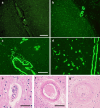
Fig. 1
Fig. 1
Morphologies of arteriolosclerosis and Cerebral amyloid angiopathy (CAA) with different scores in AD brains. CAA score was assessed using thioflavin-S fluorescence (green). a Sparse amyloid presence in both leptomeningeal and cortical vessels was rated as score 1. b Strong, circumferential amyloid deposition in some, but not all vessels were rated as 2. c Widespread, strong amyloid deposition in both leptomeningeal and cortical vessels were rated as score 3. d The most severe cases, exceeding the criteria for score 3 were rated as 4. Arteriolosclerosis score was assessed using hematoxylin and eosin-staining. e Cases with mild arteriolosclerosis, characterized by mild fibrosis and slight medial thickening were rated as 1. f Cases with moderate arteriolosclerosis, with evident fibrosis and moderate medial thickening were rated as 2. g Cases with severe arteriolosclerosis, marked by extensive fibrosis and pronounced medial thickening were rated as 3. Scale bars: 200 µm (A, B); 100 µm (C, D); 40 µm (E); 50 µm (F, G)
Fig. 2
Fig. 2
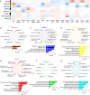
Associations of APOE and demographic/pathological variables with lipid class sums. a Pie chart showing the relative abundances of each lipid class sum. Pie charts showing the relative abundances of each lipid class sum across APOE genotypes. b Associations of demographic factors, APOE genotype, and vascular pathology with alterations in lipid class sums. The regression coefficient (β) and the corresponding p-value shown in parentheses were derived from multivariable linear regression models. p-values < 0.05 are displayed. CAA cerebral amyloid angiopathy; ARS arteriolosclerosis; LV large vessels; SV small vessels
Fig. 3
Fig. 3
Lipid modules linked to APOE and demographic/pathological variables. a Module-trait relationships between module eigengenes (MEs) and sample characteristics are shown. The correlation coefficient (r) and the corresponding p-value in the parentheses are indicated in each module. p-values < 0.05 are displayed. WGCNA results from the lipidomics dataset, highlighting modules associated with APOE genotype and demographic factors (b-g). Top‐ranked hub lipids and GO terms through LION with the highest intramodular connectivity in module brown (b), blue (c), yellow (d), red (e), green and (f), turquoise (g) are shown. CAA cerebral amyloid angiopathy; ARS arteriolosclerosis; LV large vessels; SV small vessels; WGCNA weighted correlation network analysis; GO gene ontology; LION lipid ontology
Fig. 4
Fig. 4
Associations of APOE with lipid acyl chain composition. Two-dimensional lipid class plot showing the associations of APOE2 or APOE4 allelic numbers with lipid acyl chain composition. The x-axis represents the total number of carbon atoms in acyl chain (Total C), and the y-axis represents the total number of double bonds (Total DB). Correlation strength and direction are depicted by the color scale within each grid cell. *p < 0.05. a PE Phosphatidylethanolamine, b PE-P Phosphatidylethanolamine-plasmalogens, c PC Phosphatidylcholine, d PS Phosphatidylserine, e SM Sphingomyelin, f LPE Lysophosphatidylethanolamine, g PI Phosphatidylinositol, h PG Phosphatidylglycerol, BMP bis(monoacylglycero)phosphate, i CER Ceramide, j LPC Lysophosphatidylcholine, k CAR Acyl-carnitine
Fig. 5
Fig. 5
Aβ modulates sphingolipid metabolism in human iPSC-derived vascular mural-like cells. a Representative immunostaining images of α-SMA in isogenic human iPSC-derived vascular mural-like cells (iVMLCs) with APOE ε3/ε3 (APOE3) or ε4/ε4 (APOE4). b The mRNA expressions of vascular mural cell markers (ACTA2 and PDGFRβ) and endothelial cell markers (PECAM1 and OCLN) were quantified by RT-qPCR in iVMLCs (ε3/ε3 or ε4/ε4), human brain vascular smooth muscle cells (HBSMC, ε3/ε4), and human cerebral microvascular endothelial cell line (hCMEC/D3, ε3/ε3), followed by normalization to ACTB mRNA expression. b, d The iVMLCs were treated with Aβ40 (10 µM), Aβ42 (5 µM), or vehicle (0.25% DMSO) for 48 h. Cell viability and proliferation were assessed using MTT (c) and BrdU (d) assays, respectively. e The cell lysates were analyzed for neutral sphingomyelinase (nSMase) activity. f, g Amounts of sphingomyelin (f; SM) and ceramide (g; CER) in the iVMLCs were measured by ELISA and normalized to protein concentrations. Data are presented as mean ± SEM (n = 3–4 technical replicate/each). Scale bars = 100 µm. *p < 0.05, **p < 0.01, ***p < 0.001, and ****p < 0.0001 by one-way ANOVA followed by Tukey’s post hoc test

References

    1. Abbott NJ, Friedman A (2012) Overview and introduction: the blood-brain barrier in health and disease. Epilepsia 53(Suppl 6):1–6. 10.1111/j.1528-1167.2012.03696.x - PMC - PubMed
    1. Ali H, Kobayashi M, Morito K, Hasi RY, Aihara M, Hayashi J et al (2023) Peroxisomes attenuate cytotoxicity of very long-chain fatty acids. Biochim Biophys Acta Mol Cell Biol Lipids 1868:159259. 10.1016/j.bbalip.2022.159259 - PubMed
    1. Antonny B, Vanni S, Shindou H, Ferreira T (2015) From zero to six double bonds: phospholipid unsaturation and organelle function. Trends Cell Biol 25:427–436. 10.1016/j.tcb.2015.03.004 - PubMed
    1. Bandaru VV, Troncoso J, Wheeler D, Pletnikova O, Wang J, Conant K et al (2009) ApoE4 disrupts sterol and sphingolipid metabolism in Alzheimer’s but not normal brain. Neurobiol Aging 30:591–599. 10.1016/j.neurobiolaging.2007.07.024 - PMC - PubMed
    1. Barelli H, Antonny B (2016) Lipid unsaturation and organelle dynamics. Curr Opin Cell Biol 41:25–32. 10.1016/j.ceb.2016.03.012 - PubMed

LinkOut - more resources