. 2025 Oct 8;15(1):35079.
doi: 10.1038/s41598-025-18965-0.

The prognostic importance of left ventricular diastolic function in primary mitral regurgitation and its relation to structural changes by CMR

Affiliations

The prognostic importance of left ventricular diastolic function in primary mitral regurgitation and its relation to structural changes by CMR

Hossam Lababidi et al. Sci Rep. .

Abstract

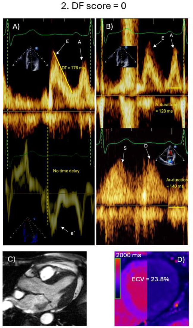
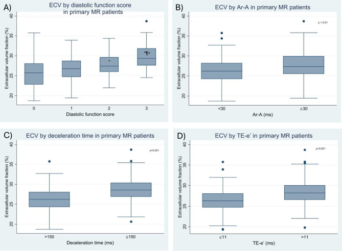
Patients with significant primary mitral regurgitation (MR) can develop left ventricular (LV) diastolic dysfunction (DD). We sought to study LV structural correlates, and prognostic significance of DD. Patients with ≥ moderate primary MR with echocardiographic and CMR scans within a median time of 5 days were included. LV volumes, EF, MR volume and fraction, presence of scar, and extracellular volume (ECV) were assessed by CMR. LV diastolic function (DF) was assessed by echocardiography using time delay between onset of mitral E velocity and mitral annulus e' velocity, deceleration time of mitral E velocity, and duration difference between pulmonary vein atrial velocity and mitral A velocity. DF score was computed based on these 3 variables. Event rate (heart failure hospitalizations, cardiovascular death, or mitral valve surgery) was higher in patients with DD versus patients with normal DF (P < 0.05). MR volumes, and ECV were significantly higher in patients with DD (P < 0.001). In multivariable cox proportional hazard model, inclusion of DF score increased AUC from 0.83 to 0.86. Thus, DD is associated with ECV, MR severity, and clinical outcomes. DD score provides incremental value for the prediction of adverse outcomes when added to clinical characteristics, MR severity, and LV end diastolic volume index.

Keywords: Diastolic function; Left ventricle; Mitral regurgitation; Outcome.


Conflict of interest statement

Competing interests: The authors declare no competing interests.

Figures

Fig. 1
Fig. 1
Echocardiographic and CMR findings in a 66-year-old female with primary MR and normal diastolic function. The Echocardiogram shows early mitral velocity (E) of 100.0 cm/s, late mitral velocity (A) of 83.2 cm/s, and E/A ratio of 1.2. No time delay is present between onset of E velocity and onset of mitral annulus septal e’ velocity. Deceleration time is 176 ms (A). Mitral A-duration is 128 ms, while pulmonary vein atrial flow (Ar) duration is 140 ms and Ar-A = 12 ms (B). By CMR (1.5 T), the MR jet is visualized in the horizontal long axis view (C). Pre-contrast T1 map is shown (D), and ECV is 23.8%. The patient was followed up for 137 months with no events.
Fig. 2
Fig. 2
Echocardiographic and CMR findings in a 67-year-old female with primary MR primary MR and advanced diastolic dysfunction. The echocardiogram showed early mitral E velocity at 119.7 cm/s, mitral A velocity at 60.0 cm/s, E/A ratio of 2, The time delay between onset of E velocity and onset of mitral annulus septal e’ velocity is 28 ms, and deceleration time is 138 ms (A). Mitral A-duration is 85 ms, while pulmonary vein atrial flow (Ar) duration is 125 ms, and Ar-A = 40 ms (B). By CMR (1.5 T), the MR jet is visualized in the vertical long axis 2-chamber view (C). Pre-contrast T1 map is shown (D), and ECV is 31.9%. During follow-up, the patient had mitral valve repair and died 12 months later.
Fig. 3
Fig. 3
Box-and-whisker plots of ECV by diastolic function score (A), Ar-A duration (B), deceleration time (C), and time delay between onset of E velocity and onset of mitral annulus e’ velocity (D). *: P < 0.05 compared to DF score 0, ¶: P < 0.05 compared to DF score 1, + , P < 0.05 compared to DF score 2.
Fig. 4
Fig. 4
Kaplan–Meier curves showing events-over-time in primary MR patients based on diastolic function score (A), and its components, Ar-A duration (B), deceleration time of mitral E velocity (C), and time delay between onset of E velocity and onset of mitral annulus e’ velocity (D). For panel (A), DF diastolic function score of 0 is shown in blue, score of 1 in purple, score of 2 in green, and score of 3 in yellow. For panel (B), Ar-A duration < 30 ms is shown in blue, and Ar-A duration ≥ 30 ms is shown in purple. For panel (C), DT > 150 ms is shown in blue, and DT ≤ 150 ms is shown in purple. For panel (D), TE-e’ ≤ 11 ms is shown in blue, and TE-e’ > 11 ms is shown in purple.

References

    1. Avierinos, J.-F. et al. Natural history of asymptomatic mitral valve prolapse in the community. Circulation106, 1355–1361 (2002). - PubMed
    1. Enriquez-Sarano, M., Akins, C. W. & Vahanian, A. Mitral regurgitation. Lancet373, 1382–1394 (2009). - PubMed
    1. Bakkestrøm, R. et al. Hemodynamic characteristics in significant symptomatic and asymptomatic primary mitral valve regurgitation at rest and during exercise. Circ Cardiovasc Imaging11, e007171 (2018). - PubMed
    1. Liu, B. et al. Myocardial fibrosis in asymptomatic and symptomatic chronic severe primary mitral regurgitation and relationship to tissue characterisation and left ventricular function on cardiovascular magnetic resonance. J. Cardiovasc. Magn. Reson.22, 86 (2020). - PMC - PubMed
    1. Edwards, N. C. et al. Quantification of left ventricular interstitial fibrosis in asymptomatic chronic primary degenerative mitral regurgitation. Circ. Cardiovasc. Imaging7, 946–953 (2014). - PubMed

LinkOut - more resources