

Development and external validation of a mixed-reality aneurysm clipping simulator
- PMID: 41062869
- PMCID: PMC12508002
- DOI: 10.1007/s10143-025-03846-x
Development and external validation of a mixed-reality aneurysm clipping simulator
Abstract
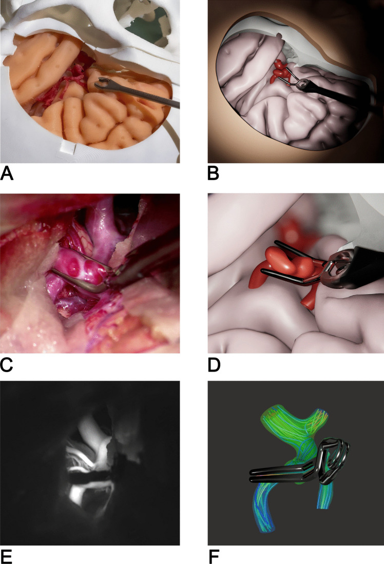
Nowadays, surgical treatment of cerebral aneurysms remains one of the most demanding disciplines in neurosurgery. The increasing shift toward endovascular interventions leads to a decline in open surgical cases. This fact leaves residents and young neurosurgeons with fewer training opportunities and limited to complex and high-risk aneurysms. There is a growing need for realistic simulation tools to enhance neurosurgical training and preoperative planning. We developed and externally validated a patient-specific mixed-reality simulator for cerebral aneurysm clipping, during the research project "Medical EDUcation in Surgical Aneurysm Clipping (MEDUSA)". Our approach combines physical phantoms of the skull and brain tissue with virtual intracranial blood vessels, including a virtual intracranial aneurysm. Real surgical instruments provide an immersive training environment featuring integrated blood flow simulation for evaluating clipping strategies. A life-sized skull with silicone brain lobes is mounted in a standard neurosurgical head clamp. Optical tracking synchronizes the position of a real clip applier and an emulated surgical microscope with the corresponding virtual environment, allowing true mixed-reality interaction. After aneurysm clipping, blood flow is automatically simulated to assess residual aneurysms or stenoses of the parental vessels. We conducted an external validation with 40 neurosurgeons at two international events. Participants completed a 32-item questionnaire evaluating face and content validity on a 5-point Likert scale. Participants' surgical experience ranged from novice to expert (> 15 years). Average ratings for simulator realism and educational value were high, with mean scores between 3.13 and 4.25. The highest ratings were for the blood flow simulation (4.25) and the simulator's potential for preoperative planning (4.20). Most participants agreed that the physical and virtual components were valuable and that the simulator should be integrated into neurosurgical training and standard surgical workflows. Our mixed-reality simulator achieved robust face and content validity among a diverse group of neurosurgeons. Combining real surgical instruments with a deformable virtual aneurysm model, including blood flow simulation, offers a high level of realism and immediate objective feedback.
Keywords: Aneurysm clipping; Blood flow simulation; Mixed-reality; Validation.
© 2025. The Author(s).
Conflict of interest statement
Declarations. Ethics approval and consent to participate: The local ethics committee (Ethikkommission der medizinischen Fakultät der Johannes Kepler Universität; EK Nr:1082/2023) approved the validation study design. All procedures performed were in accordance with the ethical standards of the institutional and national research committees, as well as with the 1964 Helsinki Declaration and its later amendments or comparable ethical standards. All participants took part voluntarily, and informed consent was waived by the ethics committee. Consent for publication: All authors have read and approved the final manuscript and consent to its publication. Competing interests: Stefan Schaffelhofer and Robert Prückl are co-owners of cortEXplore GmbH. The other authors have no relevant financial or non-financial interests to disclose. Clinical trial number: Not applicable.
Figures


References
-
- Rehder R et al (2016) The role of simulation in neurosurgery. Childs Nerv Syst 32:43–54 - PubMed
-
- Gnanalingham KK, Apostolopoulos V, Barazi S, O’Neill K (2006) The impact of the international subarachnoid aneurysm trial (ISAT) on the management of aneurysmal subarachnoid haemorrhage in a neurosurgical unit in the UK. Clin Neurol Neurosurg 108:117–123 - PubMed
-
- Wurm G, Lehner M, Tomancok B, Kleiser R, Nussbaumer K (2011) Cerebrovascular biomodeling for aneurysm surgery: simulation-based training by means of rapid prototyping technologies. Surg Innov 18:294–306 - PubMed
Publication types
MeSH terms
LinkOut - more resources
Full Text Sources
Medical
