. 2025 Oct 8;23(1):544.
doi: 10.1186/s12916-025-04378-3.

Elevated VAMP8 expression promotes cervical cancer progression by enhancing autophagy via HIF-1 pathway

Affiliations

Elevated VAMP8 expression promotes cervical cancer progression by enhancing autophagy via HIF-1 pathway

Yue Wang et al. BMC Med. .

Abstract

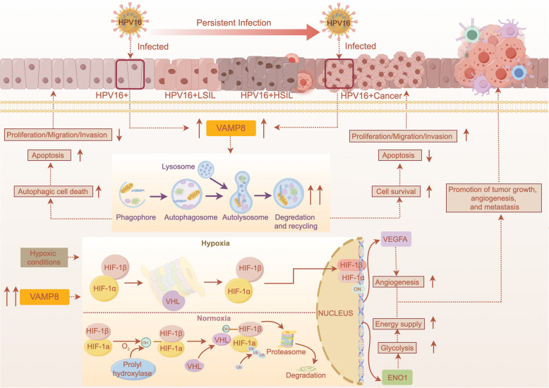
Background: Cervical cancer, prevalent in low- and middle-income countries, is primarily caused by high-risk HPV16. Vesicle-Associated Membrane Protein 8 (VAMP8), involved in vesicle trafficking and autophagy, may influence HPV16-related cervical cancer progression.

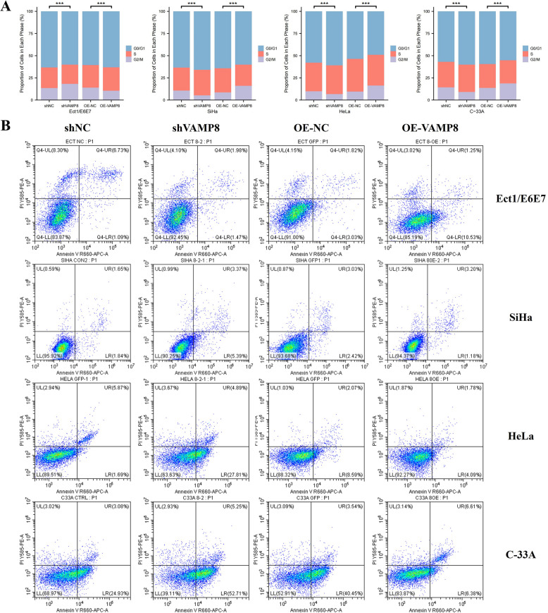
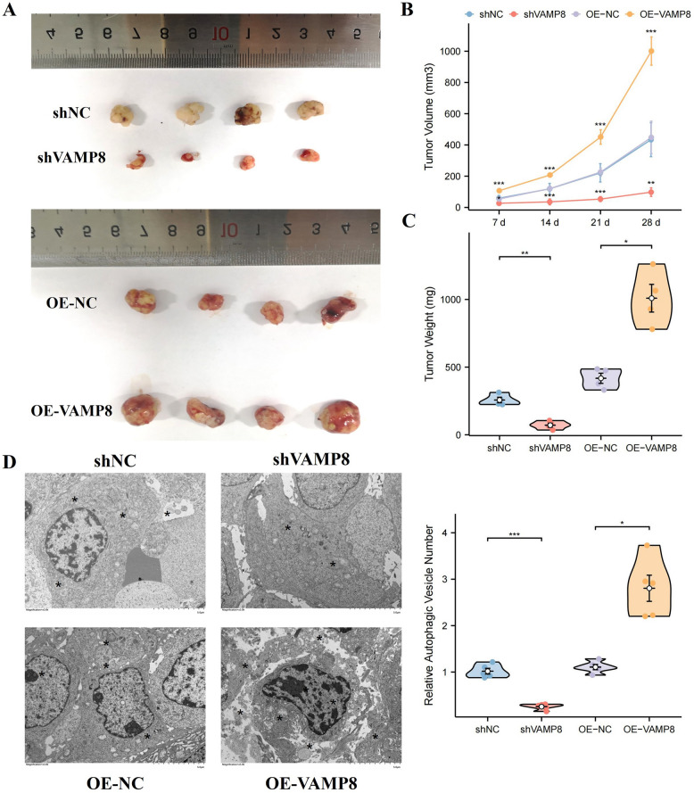
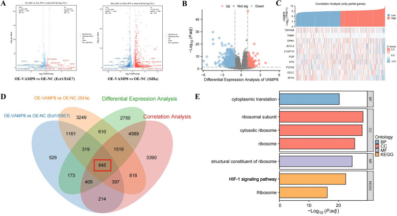
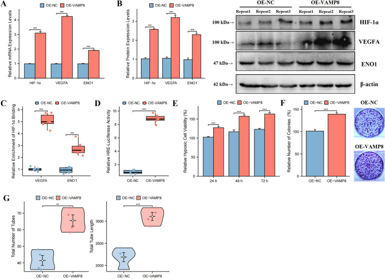
Methods: VAMP8 expression was evaluated in cervical tissue specimens from patients with HPV16-positive lesions (including low- and high-grade squamous intraepithelial lesions and cancer) and HPV-negative normal controls using proteomics, qPCR, and immunohistochemistry. A Cox proportional hazards model for prognosis was developed using immunohistochemical data from a cohort of cervical cancer patients. The clinical significance of VAMP8 was further assessed using RNA-seq and clinical data from The Cancer Genome Atlas-Cervical Cancer (TCGA-CESC) cohort. The effects of VAMP8 on autophagy and tumor progression were examined in HPV16 E6/E7-immortalized cervical epithelial cells (Ect1/E6E7) and cervical cancer cell lines (SiHa, HeLa, C-33A) in vitro, and in a SiHa xenograft model in vivo. Transcriptomic analysis of Ect1/E6E7 and SiHa cells identified VAMP8-regulated pathways. Chromatin immunoprecipitation (ChIP) and dual-luciferase reporter assays in SiHa cells were used to confirm the regulation of the HIF-1 pathway.

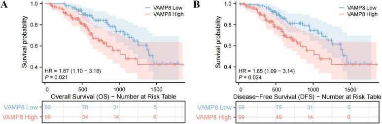
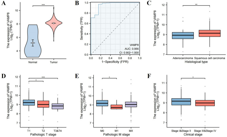
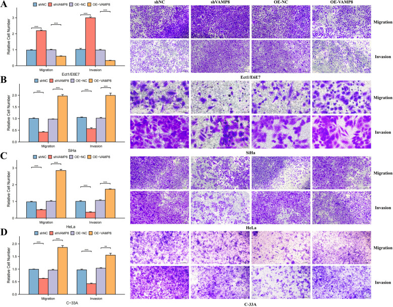
Results: VAMP8 was upregulated in HPV16-positive samples, particularly in low-grade squamous intraepithelial lesions (LSIL). Elevated VAMP8 correlated with poor survival outcomes and advanced tumor stages. VAMP8 enhanced autophagy and reduced proliferation and invasiveness in HPV16-positive cervical cells but increased in established cancer cell lines. In vivo, VAMP8 overexpression promoted tumor growth and autophagy. The HIF-1 pathway emerged as a key regulatory axis of VAMP8, enhancing hypoxic responses and angiogenesis.

Conclusion: Elevated VAMP8 in HPV16-associated cervical cancer promotes tumor progression by enhancing autophagy via the HIF-1 pathway, suggesting its potential as a diagnostic and prognostic biomarker.

Keywords: Autophagy; Biomarker; Cervical cancer; HIF-1 pathway; HPV16; VAMP8.


Conflict of interest statement

Declarations. Ethics approval and consent to participate: This study was conducted in full accordance with the Declaration of Helsinki and was approved by the Institutional Review Board of the Obstetrics and Gynecology Hospital of Fudan University for the use of human clinical specimens (Approval No. 2019–53). All participants provided written informed consent prior to their inclusion in the study. All animal experiments were performed in compliance with institutional guidelines and were reviewed and approved by the Animal Care and Use Committee of the Obstetrics and Gynecology Hospital of Fudan University. The study is reported in accordance with the ARRIVE guidelines for animal experiments and the TRIPOD statement for the prognostic prediction study. Consent for publication: Not applicable. Competing interests: The authors declare no competing interests.

Figures

Fig. 1
Fig. 1
VAMP8 Expression analysis in cervical tissues and cells. A, B Proteomic analysis (A) and qPCR (B) of VAMP8 in HPV16-positive and HPV16-negative tissues. C qPCR measurement of VAMP8 mRNA levels in cervical cell lines. D Immunohistochemical staining of VAMP8 in postoperative specimens
Fig. 2
Fig. 2
Prognostic analysis of low/high VAMP8 expression based on immunohistochemical staining of postoperative cervical cancer specimens. A, B Kaplan–Meier curves for overall survival (OS) (A) and disease-free survival (DFS) (B) in patients with low/high VAMP8 expression
Fig. 3
Fig. 3
Analysis of VAMP8 expression in cervical cancer tissues and its clinical correlation based on the TCGA-CESC database. A Comparison of VAMP8 expression between normal and tumor tissues. B Receiver operating characteristic (ROC) curve illustrating the diagnostic performance of VAMP8. C VAMP8 expression across different histological subtypes (adenocarcinoma vs. squamous cell carcinoma). D-F VAMP8 expression in relation to pathological T stage (D), pathological M stage (E), and clinical stage (F)
Fig. 4
Fig. 4
Correlation analysis between VAMP8 expression and immune cell infiltration in cervical cancer. A Correlation between VAMP8 expression and various immune cell types. The size and color of the circles represent the strength of correlation and significance, respectively. B Boxplots comparing immune cell enrichment scores for DC, aDC, Tcm, and NK cells between high and low VAMP8 expression groups. C, D Scatter plots illustrating the relationship between VAMP8 expression and the enrichment of DCs and NK cells
Fig. 5
Fig. 5
VAMP8 modulation and autophagic vesicles in cervical cell lines. A qPCR analysis confirming VAMP8 knockdown and overexpression in Ect1/E6E7, SiHa, HeLa, and C-33A cell lines. shNC: negative control for knockdown; shVAMP8: VAMP8 knockdown; OE-NC: negative control for overexpression; OE-VAMP8: VAMP8 overexpression. B Transmission electron microscopy images of autophagic vesicles in VAMP8 knockdown and overexpression cell lines across the four cervical cell lines. Scale bar = 2 μm
Fig. 6
Fig. 6
Effects of VAMP8 knockdown and overexpression on cell proliferation measured by CCK-8 assay. A–D Cell proliferation was assessed at 0, 24, 48, and 72 h in Ect1/E6E7, SiHa, HeLa, and C-33A cell lines, respectively
Fig. 7
Fig. 7
Effects of VAMP8 knockdown and overexpression on cell migration and invasion in cervical cell lines. A-D Transwell migration and invasion assays were performed in Ect1/E6E7, SiHa, HeLa, and C-33A cell lines, with relative cell numbers quantified
Fig. 8
Fig. 8
Flow cytometry analysis of cell cycle distribution and apoptosis in cervical cell lines. A Cell cycle distribution analysis after VAMP8 knockdown or overexpression. B Annexin V-APC/PI staining for apoptosis detection with VAMP8 knockdown or overexpression
Fig. 9
Fig. 9
Effects of VAMP8 on tumor growth and autophagy in vivo. A Representative images of excised tumors from nude mice (n = 4 per group) at day 28 post-injection. B Tumor volume growth curve measured over 28 days. C Violin plots showing tumor weight comparison across groups. D Transmission electron microscopy images of tumor tissues displaying autophagic vesicles in different experimental conditions
Fig. 10
Fig. 10
Analysis of VAMP8-regulated genes and enriched pathways in cervical cancer. A Volcano plots showing differentially expressed genes between VAMP8-overexpressing and control Ect1/E6E7 and SiHa cells. B Volcano plot of single-gene differential expression analysis for VAMP8 in the TCGA-CESC dataset. C Heatmap showing the correlation analysis between VAMP8 expression and other genes in the TCGA-CESC dataset. D Venn diagram depicting the overlap of genes identified through differential expression and correlation analyses across the two cell lines and TCGA data. E GO and KEGG enrichment analysis of the common genes, highlighting key biological processes and pathways
Fig. 11
Fig. 11
Effects of VAMP8 overexpression on HIF-1 signaling, cell viability, and angiogenesis in SiHa cells. A Relative mRNA expression levels of HIF-1α, VEGFA, and ENO1 in VAMP8-overexpressing cells compared to controls. B Relative protein expression levels of HIF-1α, VEGFA, and ENO1 in VAMP8-overexpressing cells. C ChIP assay showing the relative enrichment of HIF-1α binding to the VEGFA and ENO1 promoters. D Luciferase reporter assay of HRE-linked activity in VAMP8-overexpressing cells. E Relative hypoxic cell viability at 24 h, 48 h, and 72 h. F Colony formation assay showing the relative number of colonies formed under hypoxia. G HUVEC tube formation assay displaying the total number of tubes and total tube length formed in the presence of conditioned media from VAMP8-overexpressing cells
Fig. 12
Fig. 12
Schematic representation of VAMP8’s role in HPV16-associated cervical cancer progression

References

    1. Hull R, Mbele M, Makhafola T, Hicks C, Wang SM, Reis RM, et al. Cervical cancer in low and middle-income countries. Oncol Lett. 2020;20(3):2058–74. - PMC - PubMed
    1. LaVigne AW, Triedman SA, Randall TC, Trimble EL, Viswanathan AN. Cervical cancer in low and middle income countries: addressing barriers to radiotherapy delivery. Gynecol Oncol Rep. 2017;22:16–20. - PMC - PubMed
    1. Zhang S, Xu H, Zhang L, Qiao Y. Cervical cancer: Epidemiology, risk factors and screening. Chin J Cancer Res. 2020;32(6):720–8. - PMC - PubMed
    1. Choi S, Ismail A, Pappas-Gogos G, Boussios S. HPV and cervical cancer: a review of epidemiology and screening uptake in the UK. Pathogens (Basel, Switzerland). 2023;12(2). 10.3390/pathogens12020298. - PMC - PubMed
    1. Okunade KS. Human papillomavirus and cervical cancer. J Obstet Gynaecol. 2020;40(5):602–8. - PMC - PubMed

MeSH terms

Substances