

Analysis of ultrastructural defects in sperm by transmission electron microscopy in asthenozoospermia patients: a study from multiple centers across China
- PMID: 41063321
- PMCID: PMC12506306
- DOI: 10.1186/s40364-025-00841-8
Analysis of ultrastructural defects in sperm by transmission electron microscopy in asthenozoospermia patients: a study from multiple centers across China
Abstract
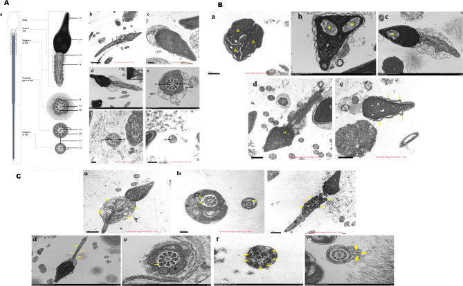
This study aimed to identify the ultrastructural features of sperm in Chinese asthenozoospermia patients and to evaluate their clinical implications. A total of 139 individuals, including 106 asthenozoospermia patients and 33 fertile controls, were recruited from multiple centers across China. Sperm ultrastructural defects were examined using transmission electron microscopy (TEM), while conventional sperm quality was assessed by light microscopy. Compared with the control group, the asthenozoospermia group showed significant ultrastructural defects, particularly in the sperm tail, including mitochondrial and axonemal defects. Based on tail ultrastructure, participants were further categorized into four groups (normal ultrastructure, simple abnormal axonemes, simple abnormal mitochondria, and both abnormality in axonemes and mitochondria). All three abnormal groups indicated significantly lower value in sperm (motile/kinematic) quality compared to the normal group. However, no statistically significant differences in sperm quality were observed among the three abnormal groups. These nationwide findings indicated that TEM could remedy the limitations of conventional light microscopy, which is difficult to localize organelle-specific defects. This capability may ultimately inform individualized diagnostic and therapeutic strategies.
Keywords: Asthenozoospermia patient; Sperm ultrastructure; Transmission electron microscopy.
© 2025. The Author(s).
Conflict of interest statement
Declarations. Ethics approval and consent to participate: This study was approved by Ethics Committee (No. LLPJ2022002). The purpose of this study was explained to each patient, and written informed consent was obtained from all subjects prior to enrolment. Consent for publication: Not applicable. Competing interests: The authors declare no competing interests.
Figures

References
-
- Ortega C, Verheyen G, Raick D, Camus M, Devroey P, Tournaye H. Absolute asthenozoospermia and ICSI: what are the options? Hum Reprod Update. 2011;17:684–92. - PubMed
-
- Mobberley MA. Electron microscopy in the investigation of asthenozoospermia. Br J Biomed Sci. 2010;67:92–100. - PubMed
-
- Li WN, Zhu L, Jia MM, Yin SL, Lu GX, Liu G. Missense mutation in DNAJB13 gene correlated with male fertility in asthenozoospermia. Andrology. 2020;8:299–306. - PubMed
-
- Nowicka-Bauer K, Lepczynski A, Ozgo M, Kamieniczna M, Fraczek M, Stanski L et al. Sperm mitochondrial dysfunction and oxidative stress as possible reasons for isolated asthenozoospermia. J Physiol Pharmacology: Official J Pol Physiological Soc. 2018;69:403-417. - PubMed
-
- Moretti E, Sutera G, Collodel G. The importance of transmission electron microscopy analysis of spermatozoa: diagnostic applications and basic research. Syst Biology Reproductive Med. 2016;62:171–83. - PubMed
Publication types
Grants and funding
LinkOut - more resources
Full Text Sources
