Review
. 2025 Sep 23:13:1631563.
doi: 10.3389/fped.2025.1631563. eCollection 2025.

Breathe easy, baby, breathe. Lung ultrasound in neonatal critical care

Affiliations
Review

Breathe easy, baby, breathe. Lung ultrasound in neonatal critical care

Mateusz Jagła et al. Front Pediatr. .

Abstract

Lung ultrasound (LUS) has emerged as an essential tool in neonatology over the past two decades, offering unique advantages for this patient population. The small size, high water content, and delayed rib calcification of neonates make them particularly suited for ultrasonographic imaging. By replacing traditional chest radiographs, it significantly reduces exposure to ionizing radiation. Furthermore, it is widely accessible, easy to use, and provides repeatable, real-time imaging without requiring patient transport. These features make it invaluable in managing acute respiratory conditions, where timely intervention is critical. This review emphasizes the role of LUS in neonates with acute respiratory distress as a fundamental component of the point-of-care ultrasound (PoCUS) protocol. The technique is crucial for conditions such as respiratory distress syndrome (RDS), supporting decisions on surfactant therapy. It also aids in diagnosing and managing air-leak syndromes like pneumothorax (PTX) and detecting congenital respiratory malformations. Additionally, LUS ensures safer transport of critically ill neonates and optimizes mechanical ventilation. By delivering accurate, real-time imaging, LUS has become an essential diagnostic tool in infant care. Its integration into clinical practice enhances the management of life-threatening conditions, making it an essential skill for clinicians in neonatal intensive care units (NICU) and during neonatal transport.

Keywords: acute respiratory failure; lung ultrasound; neonatal transport; neonate; point-of care ultrasound.


Conflict of interest statement

The authors declare that the research was conducted in the absence of any commercial or financial relationships that could be construed as a potential conflict of interest.

Figures
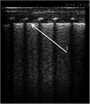
Figure 1
Figure 1
Universal alveolar-interstitial pattern (“white lung”). The female infant was delivered by Caesarean section at 27 weeks of gestation, with a birth weight of 1,150 grams. Her Apgar scores were 1, 2, 4, and 5, and she did not receive prenatal corticosteroids. Mechanical ventilation was initiated after delivery. First day of life. (1) Rough appearance of the pleural line. Linear probe, longitudinal scan.
Figure 2
Figure 2
Diffuse alveolar-interstitial pattern (“white lung”) with subpleural thin consolidations. (1) Occurs in severe RDS. Linear probe, longitudinal scan. Same patient as Figure 1.
Figure 3
Figure 3
Tension pneumothorax of the right pleural cavity. A full-term neonate, two days of age, diagnosed with congenital pneumonia, was initiated on nasal continuous positive airway pressure (nCPAP) two hours prior. (1) Thymus—mediastinum displaced to the left side. (2) Ossification nucleus in the sternum. (3) free air chamber in the pleural cavity on the right side.
Figure 4
Figure 4
Pneumothorax. Same patient as Figure 3. (1) Pneumothorax in M-mode option—“barcode” or “stratosphere” image. (2) Pneumothorax in 2D presentation.
Figure 5
Figure 5
Lung point in pneumothorax. A 32-week premature infant was resuscitated at birth with an Ambu bag. (1) Lung point. (2) Free air chamber in the right pleural cavity. (3) Confluent B lines in the aerated lung.
Figure 6
Figure 6
Fluid detected in the left pleural cavity, longitudinal lung ultrasound. The patient is a 10-day-old newborn with coarctation of the aorta following cardiac surgery involving repair of the aortic arch. (1) Large volume of fluid present in the left pleural cavity. (2) Complete atelectasis of the lower lobe of the left lung.
Figure 7
Figure 7
(A) CXR. Left-sided congenital diaphragmatic hernia. (B) LUS. Left side of sternum lacks pleural line, polycyclic echoes corresponding to intestinal echostructure (1). Transversal position of the linear probe. A female infant was delivered via C-section at 39 weeks, weighing 3,000 g with Apgar scores of 4/6/6/7. She required immediate intubation, SIMV with 100% O2, surfactant therapy, and presented with severe pulmonary hypertension, systemic hypotension, and heart failure.
Figure 8
Figure 8
(A) CXR. Right-sided CPAM, type III on histological examination. Heterogeneous right lung distension causing mediastinal shift to the left side (1. endotracheal tube position), with signs of pressure on the left lung. (B) LUS. Right lung distention—a fluid cystic lesion in the medial upper part of the right lung displaced to the left side relative to the sternum. Emphysematous bulla (2). Fluid cystic lesion (3). Longitudinal position of the linear probe. (C) LUS. Fluid cystic lesion in the lower part of the right lung. Longitudinal position of the linear probe. A male infant, delivered by caesarean section at 31 weeks (birth weight: 1950 g) with prenatally diagnosed CPAM and polyhydramnios, required intubation, repeated surfactant administration, and mechanical ventilation postnatally. Despite high-frequency oscillatory ventilation (FiO2 1.0), nitric oxide, vasopressors, and epoprostenol for PPHN and hypotension, the left lung remained abnormal after surgery without expansion. The patient died.
Figure 9
Figure 9
(A) LUS. Intrapulmonary sequestration—atelectatic area with heterogeneous structure, mostly solid (1). Transversal position of the linear probe, supradiaphragmatic region of the posterior field of the left lung. (B) LUS. The vessels marked with color Doppler are branches of the aorta. (C) Angio CT. The lesion (2). Aortic branch (3). The male neonate was admitted with respiratory failure (nCPAP, FIO2 0.4, RDS) following caesarean section at 37 weeks gestation, with a birth weight of 3,320 g and an Apgar score of 9. The mother received lamotrigine and lacosamide for epilepsy during pregnancy. Prenatal imaging indicated possible pulmonary sequestration. Echocardiography revealed an atrial septal defect and a right subclavian artery arising from the aortic arch. The respiratory disorder resolved, and the patient was discharged with a plan for outpatient follow-up at a surgical clinic in 2–3 months to assess and schedule resection of the pulmonary sequestration.

References

    1. Lichtenstein DA, Mezière GA. Relevance of lung ultrasound in the diagnosis of acute respiratory failure: the BLUE protocol. Chest. (2008) 134(1):117–25. 10.1378/chest.07-2800 - DOI - PMC - PubMed
    1. Lichtenstein DA, Mauriat P. Lung ultrasound in the critically ill neonate. Curr Pediatr Rev. (2012) 8(3):217–23. 10.2174/157339612802139389 - DOI - PMC - PubMed
    1. Jagła M, Krzeczek O, Buczyńska A, Zakrzewska Z, Kwinta P. The safety of pulmonary ultrasonography in the neonatal intensive care unit. Dev Period Med. (2018) 22(1):75. 10.34763/devperiodmed.20182201.7580 - DOI - PMC - PubMed
    1. Jagła M, Grudzień A, Starzec K, Tomasik T, Zasada M, Kwinta P. Lung ultrasound in the diagnosis of neonatal respiratory failure prior to patient transport. J Clin Ultrasound. (2019) 47(9):518–25. 10.1002/jcu.22766 - DOI - PubMed
    1. Fei Q, Lin Y, Yuan TM. Lung ultrasound, a better choice for neonatal pneumothorax: a systematic review and meta-analysis. Ultrasound Med Biol. (2021) 47(3):359–69. 10.1016/j.ultrasmedbio.2020.11.011 - DOI - PubMed

LinkOut - more resources