Review
. 2025 Oct 9;24(1):251.
doi: 10.1186/s12943-025-02463-y.

Advances in RNA-based cancer therapeutics: pre-clinical and clinical implications

Affiliations
Review

Advances in RNA-based cancer therapeutics: pre-clinical and clinical implications

Yubo Yan et al. Mol Cancer. .

Abstract

Cancer therapy has been revolutionised by the emergence of RNA-based therapeutics, providing several strategies and mechanisms to regulate gene expression via messenger RNA (mRNA), small interfering RNA (siRNA), microRNAs (miRNA), antisense oligonucleotides (ASOs), and RNA aptamers. The present review highlights the recent advances in the preclinical development and clinical applications of RNA-based therapeutics, focusing on the delivery strategies, biological targets, and pharmacological optimisation, together with key clinical data. mRNA therapeutics, especially those adapted from vaccine platforms are being developed for the cancer immunotherapy and protein replacement, while siRNAs and ASOs enable highly specific gene silencing and splice correction. miRNA therapies show potential for diverse oncogenic pathway control, despite ongoing challenges in the delivery and specificity. RNA aptamers are obtaining attention as tumor-targeting agents in the drug delivery systems. Progress in lipid nanoparticles, chemical modifications, and tissue-specific delivery has improved the stability and efficacy of these agents. Early-phase clinical trials report encouraging outcomes in both solid tumours and haematologic malignancies, particularly in overcoming resistance and modulating the tumor microenvironment (TME). Although challenges remain in scalability, immune activation, and deep-tumour penetration, RNA-based strategies are advancing towards integration into clinical oncology. Continued refinement of delivery technologies and targeted trial designs will be critical for translating these therapies into effective, personalized cancer treatments.

Keywords: Antisense oligonucleotides; Cancer immunotherapy; MRNA vaccine; RNA aptamers; SiRNA.


Conflict of interest statement

Declarations. Ethics approval and consent to participate: Not applicable. Consent for publication: Not applicable. Competing interests: The authors declare no competing interests.

Figures

Fig. 1
Fig. 1
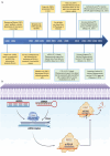
A The advancement of mRNA-based therapeutics has progressed through distinct phases. From 1961 to 1990, foundational discoveries laid the groundwork, including the identification of mRNA and early delivery techniques Such as Liposomes. Between 1990 And 2019, the focus shifted to experimental applications in vaccines, immunotherapy, and gene editing, highlighted by key milestones such as the first clinical trials using mRNA-transfected dendritic cells (DCs) and improvements in LNP delivery systems. The COVID-19 pandemic (2019–present) marked a pivotal moment, bringing global recognition to mRNA technology with the rapid rollout of vaccines such as mRNA-1273 and BNT162b. This evolution highlights the platform’s versatility, rapid development capabilities, and broad therapeutic potential in treating infectious diseases, cancer, and genetic conditions. Inpisrted and redrawn from Ref [10]. B Conventional mRNA constructs consist of a 5’ cap, 5’ UTR, gene of interest (GOI), 3’ UTR, and a poly(A) tail. Upon reaching the cytoplasm, this mRNA is directly translated into the target protein. In contrast, saRNA includes additional viral-derived elements, such as non-structural proteins (nsP1–4) and a subgenomic promoter (SGP). After cellular entry, saRNA first produces nsP1–4, which assemble into a replication complex that synthesizes complementary negative-sense RNA. This complex subsequently transitions into a replicase that generates multiple saRNA copies and subgenomic RNAs. These subgenomic RNAs are then efficiently translated, enabling sustained and amplified protein production compared to conventional mRNA. Inpisrted and redrawn from Ref [11]. (Created with Biorender.com)
Fig. 2
Fig. 2
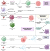
This figure presents the diverse strategies employed in mRNA-based cancer therapy, emphasizing key mechanisms and clinical outcomes. DC vaccines encoding tumor-associated antigens such as CEA and KRAS G12V stimulate strong T-cell responses, contributing to tumor regression and extended Survival. mRNA modifications, Such as 3′-end extensions, improve transcript stability and enhance the expression of pro-apoptotic proteins like apoptin for selective tumor cell killing. Combination therapies, for example survivin mRNA paired with STAT3 inhibitors, help reshape the immunosuppressive TME. Adoptive T-cell therapies using mRNA-electroporated chimeric antigen receptors (CARs) deliver transient but powerful antitumor activity. However, limited vaccine efficacy in hematologic malignancies highlights the need for tailored strategies in immunocompromised patients. Synthetic lethality approaches, such as targeting PELO-HBS1L in SKI-deficient tumors, and promising clinical outcomes in PCa, highlight the adaptability of mRNA platforms in oncology. Despite these advances, challenges remain, Such as the short-lived nature of CAR T-cell effects, inconsistent immune responses in blood cancers, and the suppressive TME. Additional obstacles include complex Manufacturing processes, selecting optimal antigens, and developing personalized neoantigen vaccines. Future directions should prioritize enhancing mRNA stability through advanced 3′ modifications, boosting TME infiltration via checkpoint inhibitors or cytokines, and expanding clinical trials to validate efficacy across cancer types. Strategies such as booster dosing, synthetic lethality targeting, and customized approaches for patients with impaired immunity may further optimize the potential of this evolving therapeutic platform. (Created with biorender.com)
Fig. 3
Fig. 3
A From Discovery to Regulatory Approval: The Timeline of Small Interfering RNA (siRNA). GalNAc, N-acetylgalactosamine; RISC, RNA-induced silencing complex; RNAi, RNA interference; US FDA, US Food and Drug Administration. Reprinted with permission from Wiley [43]. B The mechanism of action of siRNA in the suppression of gene expression (Created with Biorender.com)
Fig. 4
Fig. 4
Mechanisms of siRNA-Based Therapeutics in Cancer Therapy. The downregulation of TPX2, EphA2, Wnt2B, RhoA/RhoC, survivin, and RRM1/RRM2/PLK1 by siRNA can effectively suppress tumor cell proliferation. Additionally, siRNA-induced gene silencing can cause cell cycle arrest in the S phase and induce a G2/M phase shift. Increased apoptosis can also be achieved through siRNA. The downregulation of Wnt2B and RhoA/RhoC by siRNA can inhibit the EMT/PI3K/AKT axis, thereby disrupting tumor metastasis. Combining miR-520d-3p with EphA2 siRNA can exert a synergistic effect in suppressing tumor invasion. Moreover, the use of EGFR and Wnt2B siRNA can enhance sensitivity to chemotherapy. Application of PARC, as photoactivated siRNA promotes PD-L1 downregulation to increase T cell infiltration in cancer therapy. (Created with biorender.com)
Fig. 5
Fig. 5
The Essential Role of miRNAs in Cancer Therapy. miRNAs are central to cancer biology, acting as both oncogenes and tumor suppressors by regulating crucial processes such as metastasis, angiogenesis, and EMT. Pro-metastatic miRNAs, such as miR-373 and miR-10b, promote tumor invasion by downregulating targets such as CD44 and HOXD10, respectively. In contrast, tumor-suppressive miRNAs like miR-34a and the miR-200 family inhibit EMT and reduce cancer stemness by targeting CD44 and ZEB1. Circulating miRNAs, including miR-7 and miR-429, serve as highly specific and sensitive biomarkers, aiding in accurate tumor classification and prognosis. Furthermore, exosomal miRNAs such as miR-1247-3p and miR-25-3p influence the TME by activating fibroblasts and enhancing vascular permeability, highlighting their key role in cancer progression and the establishment of metastatic niches. (Created with biorender.com)
Fig. 6
Fig. 6
A The therapeutic application of ASOs in cancer therapy. B ASOs can inhibit the synthesis of protein products by obstructing the addition of the 5′ cap or the poly-A tail, or by inducing alternative splicing of newly transcribed mRNA. ASOs can impede translation by obstructing ribosome binding to mRNA or by enlisting RNase, leading to RNA breakdown [87]
Fig. 7
Fig. 7
A visual roadmap summarizing emerging strategies to enhance RNA aptamer applications in oncology. Future efforts focus on improving stability, enabling smart delivery systems, developing multiplexed aptamer cocktails, integrating AI for faster discovery, and combining aptamers with cutting-edge therapeutic platforms. (Created with Biorender.com)
Fig. 8
Fig. 8
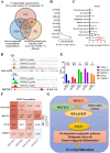
A prospective NKX2-2–mediated m6A regulation network implicating KIAA1429 in ES: A Venn diagram indicates nine transcripts in ES that are both highly expressed and positively linked with KIAA1429 expression. The nine ES-specific transcripts are prioritized according on their expression fold change. Analysis of DepMap cancer cell line data indicates a unique transcriptional profile for NKX2-2 in embryonic stem cells. Gene track data from publically accessible ES ChIP-seq datasets reveal pronounced NKX2-2 binding peaks in the promoter regions of KIAA1429 (top panel) and METTL3 (bottom panel), indicated by promoter-associated histone modifications H3K27ac and H3K4me3. (E) RNA-seq analysis following NKX2-2 knockdown showed a decrease in numerous m6A methylation-related genes. A correlation matrix demonstrates co-expression patterns among KIAA1429, METTL3, WTAP, and YTHDF3 across many ES tumor datasets. A diagram illustrating the KIAA1429-centered m6A regulation pathway and its possible influence on essential phenotypic characteristics in embryonic stem cells. *Abbreviations: ChIP – chromatin immunoprecipitation; ChIP-seq – chromatin immunoprecipitation sequencing; ES; FC – fold change. Statistical significance: *P < 0.05; **P < 0.01; ***P < 0.001. Reprinted with permission from Springer BMC Nature [104]
Fig. 9
Fig. 9
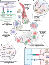
RNA changes are essential in modulating every phase of the cancer-immunity cycle, which includes the fundamental processes necessary for T cells to accurately identify and eradicate cancer cells. These alterations affect several cell types and molecular mechanisms inside the TME. The commencement of an immune response is initiated by APCs, including DCs, which process antigens and display co-stimulatory chemicals. The m6A methylation of mRNA, facilitated by METTL3, augments the production of the co-stimulatory molecule CD80, hence enhancing antigen presentation and T cell priming. B Upon activation, CTLs traverse the circulation and lymphatic system, guided by chemokines. RNA changes can modulate the expression of these chemokines. In tumor cells, METTL3 and METTL14 inhibit the transcription of CXCL9 and CXCL10 via m6A alterations, facilitating immunological exclusion in colorectal cancer. Furthermore, the enzyme PUS7, which facilitates pseudouridylation a common RNA alteration in GBM has been demonstrated to reduce CXCL10 levels. The infiltration of T cells into tumor tissue is influenced by the extracellular matrix, especially via collagen buildup. CAFs synthesize collagens such as COL10A1. In lung squamous cell carcinoma (LUSC), METTL3-mediated m6A methylation enhances the stability of COL10A1 mRNA, hence increasing its production and secretion by CAFs. Furthermore, VEGFA released by CAFs stimulates angiogenesis and can enhance METTL3 expression in NSCLC cells. CAFs produce extracellular vesicles that contain PIATs, which depend on m5C methylation to augment YBX1 protein binding, hence promoting neural remodeling in the TME. As cancer cells perish, they emit tumor antigens, including neoantigens, into the adjacent microenvironment. Tumors characterized by reduced m6A and m1A scores typically have an elevated neoantigen burden. Modifications including GPX4 m6A (facilitated by RBM15B and IGFBP2) and m5C (catalyzed by NSUN5) are associated with the activation of the STING pathway, which contributes to anti-tumor immunity. E Effective antigen presentation through MHC I molecules is required for T lymphocytes to detect tumor cells. In glioma stem cells, reduced expression of METTL3 and YTHDF2 diminishes m6A levels, which is associated with elevated MHC I expression. Concurrently, the immunosuppressive lncRNA LINC00624 stabilizes ADAR1, an enzyme responsible for adenosine-to-inosine RNA editing. This modification hinders MHC I antigen presentation and diminishes CD8+ T cell infiltration. In ICC, the m6A demethylase ALKBH5 diminishes m6A modifications on PD-L1 mRNA, enhancing its stability and facilitating immune evasion. In AML, the inhibition of the demethylase FTO reduces the production of immunological checkpoint proteins such as PD-L1, hence increasing the tumor’s vulnerability to T cell-mediated eradication. Reprinted with permission from Springer BMC Nature [178]
Fig. 10
Fig. 10
Schematics of iDR-NC/neoantigen Nanovaccines for Improved Tumor Immunotherapy. a In a unified reaction system, rolling circle replication (RCR) and rolling circle transcription (RCT) were executed concurrently, yielding concatenated CpG motifs and Stat3 shRNA strands. These nucleic acid products self-assembled into hybrid DNA-RNA microfibers (MFs) exhibiting an interwoven topology. b The resultant MFs were condensed with PPT-g-PEG, yielding iDR-NCs. The nanocomplexes were further loaded with tumor-specific neoantigens via hydrophobic interactions between the peptide antigens and the hydrophobic domains of PPT. c When administered to immunocompetent mice, the iDR-NC/neoantigen complexes specifically targeted antigen-presenting cells (APCs) within the draining lymph nodes. This elicited robust and prolonged neoantigen-specific T cell responses, resulting in efficient tumor growth suppression. Reprinted with permission from Springer Nature [226]
Fig. 11
Fig. 11
An in vitro assessment of the therapeutic effectiveness of CLPV nanoparticles (NPs) against HCC was performed. A schematic diagram depicts the therapy plan of CLPV nanoparticles for HCC. T7 endonuclease I (T7E1) tests were conducted to evaluate VEGFR2 gene editing in HepG2 and HeLa cells subjected to CLPV nanoparticles and VC liposome-mediated transfection. Western blot analysis was employed to assess VEGFR2 protein expression in HepG2 cells after treatment with liposome-transfected and nanoparticle formulations (CLP and CLPV NPs). The cytotoxic effects of various nanoparticle groups were evaluated using a CCK-8 test and compared to free paclitaxel (PTX) on SMMC-7721 and HepG2 cell lines. Data are expressed as mean ± standard deviation (SD, N = 6), with statistical significance indicated (* P < 0.05, ** P < 0.01). Quantitative RT-PCR and Western blot analyses were employed to assess IL-6 and IL-8 mRNA expression and NF-κB p65 protein levels, respectively. Data are presented as mean ± SD (N = 6), with * P < 0.05 denoting statistical significance. Western blot analysis was performed to assess the expression of NF-κB p65 and Ser276-phosphorylated proteins in HepG2 cells following treatment with PTX and NP formulations. Reprinted with permission from Elsevier [282]
Fig. 12
Fig. 12
Design And characterization of 10B/siPD-L1 nanoparticles. a A diagram illustrating the production of 10B/siPD-L1 nanoparticles and their application in the integrated approach of BNCT and immunotherapy. b ICP-MS spectra of the cRGD-PEG-PME-PBOB copolymer used for ascertaining the boron isotope composition. c Acid-base titration profile of the cRGD-PEG-PME-PBOB copolymer to evaluate its pKa. d Gel electrophoresis findings demonstrating siRNA loading efficiency at different Mass ratios of 10B polymer to siRNA. e Assessments of particle size, zeta potential, and transmission electron microscopy (TEM) Pictures of 10B/siPD-L1 nanoparticles before to and after to disulfide crosslinking. Reprinted with permission from Wiley [299]
Fig. 13
Fig. 13
Schematic diagram illustrating the synthesis pathway for the multifunctional UAAP nanoparticles and the suggested mechanism for dual inhibition of CA IX and Au-mediated improved irradiation. Reprinted with permission from Elsevier [306]
Fig. 14
Fig. 14
Depiction of MMP-2-responsive, peptide-assembled micelleplexes for improved photoimmunotherapy. Reprinted with permission from Elsevier [311]
Fig. 15
Fig. 15
ICIE combines cryosurgery with the systemic administration of cold-responsive nanoparticles (CRNPs) co-encapsulated with irinotecan (camptothecin or CPT) and PD-L1-targeting siRNA (siR) to restructure the TME. This method promotes ICD and inhibits PD-L1 expression in tumor cells, successfully converting an immunosuppressive (“cold”) TME into an immuno-active (“hot”) one. Consequently, CD8+ T lymphocytes are stimulated to facilitate both local and systemic tumor eradication. The CPT&siR CRNPs are produced utilizing a double-emulsion method, which includes CPT, siR, poly(D, L-lactide-co-glycolide) (PLGA), poly(N-isopropylacrylamide-co-butyl acrylate) (pNIPAAm-BA), chitosan-modified Pluronic F-127 (CS-PF-127), 1,2-dipalmitoyl-sn-glycero-3-phosphocholine (DPPC), and sodium chloride (NaCl). Reprinted with permission from Springer Nature [314]

References

    1. DePinho RA. The age of cancer. Nature. 2000;408(6809):248–54. - PubMed
    1. Fisher R, Pusztai L, Swanton C. Cancer heterogeneity: implications for targeted therapeutics. Br J Cancer. 2013;108(3):479–85. - PMC - PubMed
    1. Bode AM, Dong Z. Cancer prevention research — then and now. Nat Rev Cancer. 2009;9(7):508–16. - PMC - PubMed
    1. Morris KV, Mattick JS. The rise of regulatory RNA. Nat Rev Genet. 2014;15(6):423–37. - PMC - PubMed
    1. Pastor F, et al. An RNA toolbox for cancer immunotherapy. Nat Rev Drug Discovery. 2018;17(10):751–67. - PubMed

LinkOut - more resources