. 2025 Sep 30.
doi: 10.1039/d5cb00151j. Online ahead of print.

A Lyt at the end of the tunnel? Unraveling the complex interactions of the N-acetylglucosaminidase LytG in cell wall metabolism

Affiliations

A Lyt at the end of the tunnel? Unraveling the complex interactions of the N-acetylglucosaminidase LytG in cell wall metabolism

Jazmeen Hernandez et al. RSC Chem Biol. .

Abstract

The growth and division of the Gram-positive cell requires the coordinated action of enzymes involved in the synthesis and degradation of the heteropolymer peptidoglycan. Herein, we present the use of the diamide masarimycin, an inhibitor of the exo-N-acetylglucosaminidase (GlcNAcase) LytG from Bacillus subtilis, as a chemical biology probe to elucidate the biological role of this cell wall degrading enzyme. Using a combination of chemical biology and genetic approaches we provide the first evidence that LytG activity influences the elongation and division complexes in B. subtilis. Chemical inhibition of LytG resulted in dysregulated cell elongation and localization of the division plane and the induction of the cell wall stress response. In the presence of masarimycin, cells show asymmetrical thickening of the cell wall and dysregulation of division plane localization. The use of genetic and synergy/antagonism screens established connections to late-stage peptidoglycan synthesis, particularly related to cross-linking function. These results stand in stark contrast to those observed for the ΔlytG knockout, which does not exhibit these phenotypes. Cell-wall labelling with a fluorescent d-amino acid and muropeptide analysis has highlighted a functional connection between LytG, the carboxypeptidase DacA, and d,d-endopeptidases. These results highlight the use of chemical probes such as masarimycin to inform on the biological function of autolysins by providing insight into the role LytG plays in cell growth and division.


Conflict of interest statement

There are no conflicts to declare.

Figures

Fig. 1
Fig. 1. Structure of peptidoglycan showing the cleavage sites of the major autolysins in Bacillus subtilis. Inset, structure of Mas, an inhibitor of the exo-acting GlcNAcase LytG.
Fig. 2
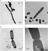
Fig. 2. Electron microscopy of B. subtilis treated with 0.75× MIC (3 μM) Mas. (A) and (B) Negative stain EM showing elongated cells and incomplete septation; (C) and (D) thin section EM showing accumulation of cell wall material at the poles of Mas treated cells.
Fig. 3
Fig. 3. Atomic force microscopy of B. subtilis control (A) and 0.75× MIC masarimycin treated (B) cells. Treatment with masarimycin results in a rough surface that correlates with the observed thickening of the cell wall in thin-section electron microscopy images.
Fig. 4
Fig. 4. Confocal fluorescence microscopy (40× objective) of B. subtilis cell wall stem-peptides metabolically labeled with HADA. Early exponentially growing B. subtilis cells (OD600 nm = 0.2) labeled with 1 mM HADA and Syto-13 in the absence (A) and (C) or presence (B) and (D) of 0.75× MIC Mas.
Fig. 5
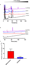
Fig. 5. (A) RP-HPLC muropeptide profile of B. subtilis in the absence or presence of Mas and comparison to the ΔlytG and ΔdacA strains. (B) RP-HPLC muropeptide profiles in the 60–120 min region. (C) Relative quantitation of relA (ppGpp) synthase as a measure of induction of stringent response upon treatment with Mas or the cell wall antibiotic cefoxitin. Experiments were run in biological and technical duplicate. Average 2−ΔΔCq with standard deviation are presented. Asterisk denotes unique muropeptides.
Fig. 6
Fig. 6. (A) Chemical genetic screen of autolysin (lytG, dacA) cell division (minC, minD, ftsH), and transpeptidases (pbpA/H/C/D) mutant strains for changes in Mas sensitivity. (B) Whole cell assay measuring exo-GlcNAcase activity in several mutant strains from the genetic screen. Results shown are the average of 2 biological and technical replicates.
Fig. 7
Fig. 7. Proposed mechanism of Mas-induced phenotypes through the inhibiton of the the exo-acting GlcNAcase LytG. Antiobiotics demonstrating synergy with masarimcyn are shown in green while antagonistic relationships are shown in red; Bac: bacitracin, Van: vancomycin, Cef: cefoxitin, Amp: ampicillin, Mas: masarimycin (A) Under normal growth conditions there is tight coordination between biosynthesis and degradation of PG. PG hydrolases such as LytG release tension in the outer layers of the cell wall releasing soluble muropeptides this allows for the migration of the essential endopeptidase LytE to migrate from the inner to the outer wall as the cell grows. This leaves the other essential endopeptidase CwlO as an integral part of the elongation and divison complexes. (B) In the presence of masarimycin, LytG is inhibited preventing the release of tension in the outermost layers of the cell wall. This prevents the outward migration of LytE as nacent PG is no longer pulled into the stress-bearing layers. This results in LytE being retained in the cell wall synthetic complex, resulting in disregulated growth and division manifesting in part by changes in cross-linking through the activity of DacA and d,d-transpeptidases (d,d-TP). Created in https://BioRender.com.

References

    1. Errington J. Aart L. T. V. Microbiology. 2020;166:425–427. - PMC - PubMed
    1. Vollmer W. Blanot D. de Pedro M. A. FEMS Microbiol. Rev. 2008;32:149–167. - PubMed
    1. Bhavsar A. P. Brown E. D. Mol. Microbiol. 2006;60:1077–1090. - PubMed
    1. Beeby M. Gumbart J. C. Roux B. Jensen G. J. Mol. Microbiol. 2013;88:664–672. - PMC - PubMed
    1. Koch A. L. Doyle R. J. J. Theor. Biol. 1985;117:137–157. - PubMed