Mesenchymal stem cell-conditioned medium accelerates type 2 diabetic wound healing by targeting TNF and chemokine signaling
- PMID: 41070349
- PMCID: PMC12504491
- DOI: 10.3389/fcell.2025.1659444
Mesenchymal stem cell-conditioned medium accelerates type 2 diabetic wound healing by targeting TNF and chemokine signaling
Abstract
Introduction: Given the crucial role of paracrine signaling in the therapeutic function of adipose tissue-derived mesenchymal stem cells (ADSCs) for skin wound repair, this study aimed to evaluate the efficacy of ADSC-conditioned medium (ACM) in enhancing type 2 diabetic (T2D) wound healing.
Methods: The effect of ACM on the viability and angiogenesis of human umbilical vein endothelial cells (HUVECs) was first evaluated using the CCK-8 assay and q-PCR analysis, respectively. Next, a T2D rat model was established through the combination of a high-fat diet and streptozotocin (STZ). Following the establishment of full-thickness skin defects in T2D rats, ACM or serum-free cultured medium was daily injected around the wound edges for 7 days. Afterward, the skin wound healing rate was analyzed, and the skin tissues were assessed by histopathological examination. The mRNA levels of TNF-α, IL-1β, IL-6, COX-2, IL-12, and IFN-γ were evaluated by q-PCR analysis. Additionally, transcriptome sequencing and immunohistochemistry were performed to reveal the potential mechanisms of ACM in T2D skin wound healing.
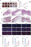
Results: ACM significantly enhanced HUVEC proliferation and angiogenesis while upregulating the expression of EGF, bFGF, VEGF, and KDR. In T2D rats, ACM accelerated wound closure and suppressed pro-inflammatory mediators (TNF-α, IL-1β, IL-6, COX-2, IL-12, and IFN-γ). Notably, transcriptome analysis revealed ACM-mediated downregulation of TNF and chemokine signaling pathways.
Discussion: ACM promotes diabetic wound healing through dual mechanisms: (1) stimulating vascularization by inducing growth factor expression and (2) modulating the inflammatory microenvironment by inhibiting TNF/chemokine cascades. These findings position ACM as a promising cell-free therapy for impaired wound healing in diabetes.
Keywords: adipose tissue-derived mesenchymal stem cells; conditioned medium; regeneration; skin wound; type 2 diabetes.
Copyright © 2025 Huang, Lin, Liu, Lin, Liao and Wu.
Conflict of interest statement
The authors declare that the research was conducted in the absence of any commercial or financial relationships that could be construed as a potential conflict of interest.
Figures


References
-
- An Y. H., Kim D. H., Lee E. J., Lee D., Park M. J., Ko J., et al. (2021). High-efficient production of adipose-derived stem cell (ADSC) secretome through maturation process and its non-scarring wound healing applications. Front. Bioeng. Biotechnol. 9, 681501. 10.3389/fbioe.2021.681501 - DOI - PMC - PubMed
-
- Carstens M. H., Quintana F. J., Calderwood S. T., Sevilla J. P., Ríos A. B., Rivera C. M., et al. (2021). Treatment of chronic diabetic foot ulcers with adipose-derived stromal vascular fraction cell injections: safety and evidence of efficacy at 1 year. Stem Cells Transl. Med. 10, 1138–1147. 10.1002/sctm.20-0497 - DOI - PMC - PubMed
