Review
. 2025 Oct 10;23(1):655.
doi: 10.1186/s12951-025-03729-9.

Constructing biomimetic microenvironments for liver regeneration

Affiliations
Review

Constructing biomimetic microenvironments for liver regeneration

Yawen Zhu et al. J Nanobiotechnology. .

Abstract

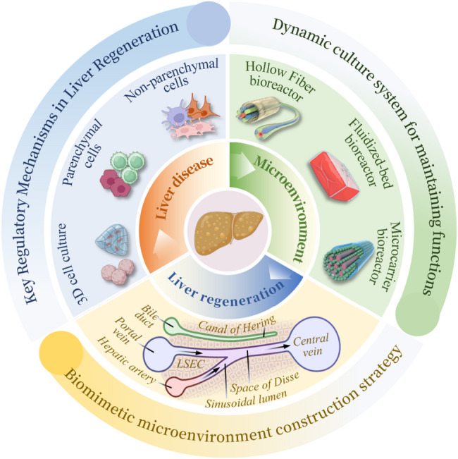
Liver regeneration is a sophisticated biological process influenced by a complex microenvironment that becomes profoundly altered in various pathological conditions. Current therapeutic approaches, including liver transplantation and pharmacological interventions, face significant limitations such as donor shortages, high costs, immune rejection, and insufficient functional recovery. Thus, alternative and innovative strategies are urgently needed. Biomimetic microenvironments constructed through tissue engineering have emerged as promising platforms, capable of recapitulating the liver's natural architecture and supporting hepatic cell functions. This review outlines key pathological features and the biological basis underlying liver regeneration, highlighting cellular plasticity, inflammation, extracellular matrix (ECM) remodeling, and immune interactions. It further discusses advanced biomimetic strategies, including 3D cell cultures, decellularized ECM hydrogels, bioprinting technologies, and dynamic culture systems like hollow fiber, fluidized-bed, and microcarrier bioreactors. These innovations facilitate accurate modeling of hepatic functions, maintain cellular differentiation, and enhance regeneration. Despite significant advancements, challenges remain in optimizing microenvironmental fidelity, ensuring clinical scalability, and translating laboratory breakthroughs into effective therapies.

Keywords: Biomaterials; Biomimetic microenvironment; Extracellular matrix; Liver diseases; Liver regeneration; Tissue engineering.


Conflict of interest statement

Declarations. Ethics approval and consent to participate: Not applicable. Consent for publication: All authors gave their consent for publication. Competing interests: The authors declare no competing interests.

Figures

Fig. 1
Fig. 1
Schematic illustration of constructing the bionic microenvironment for liver regeneration. Created by BioRender.com
Fig. 2
Fig. 2
Analysis of plasticity of cholangiocytes and hepatocytes using single-nucleus RNA sequencing of MASLD progression. (A) Immunofluorescence staining of liver tissues from normal and liver disease patients, with the square box area being a high-magnification image. Scale bars, 1,000 μm for low magnifications and 100 μm for high magnifications. (B) Immunofluorescence images of K19 or K7 and ALB double-positive cells in end-stage MASLD tissues, located around hepatocyte nodules and ducts, with yellow arrows indicating typical cells. Scale bars, 100 μm. (C) Single-nucleus RNA sequencing experimental flow chart. (D) UMAP display and cell type of samples at each stage after integration. (E) Genes marking major cell types. (F) UMAP illustration of different disease stages [83]. Copyright 2024, Springer Nature
Fig. 3
Fig. 3
Interorgan communication in ALD. Alcohol and its metabolites impair the gut barrier, allowing pathogen-associated molecules (PAMPs) to enter and activate immune responses, leading to inflammation. In ALD, levels of short-chain fatty acids (SCFAs) decrease, worsening gut permeability. Alcohol also disrupts gut microbiota, reduces beneficial bacteria like Akkermansia muciniphila, and lowers intestinal dendritic cells. It impairs FXR signaling, affecting bile acid regulation. In white adipose tissue, alcohol promotes fat breakdown, releasing fatty acids that accumulate in the liver as triglycerides, contributing to steatosis and inflammation. Multiple pathways together drive liver injury in ALD [104]. Copyright, 2024, BMJ Publishing
Fig. 4
Fig. 4
(A) Diagram illustrating TGFβ receptor inhibitor treatment. Mice with ΔMdm2 livers received oral gavage of TGFβ receptor inhibitor or vehicle twice daily. (B) Quantification of protein band intensity from immunoblot analysis. (C) Representative p21 IHC images of kidney, brain, and lung tissues of the treatment group mice. (D) Manual quantification of p21 + cells in kidney cortex, shown as average number of positive cells per field of view per mouse. (E) Artificially quantification of p21 + brain cells per mm². (F) Automatic quantification of p21 + lung cells [112]. Copyright 2024, Springer Nature. (G) H&E-stained histological sections of ileum. Scale bar, 40 μm. (H) UMAP visualization of all single cells. (I) Immunofluorescence staining of Cd3d, Cd79a, Gzmb in Peyer’s patches and intestinal epithelium, with DAPI (blue) used for nuclear counterstaining. Scale bar, 100 μm. (J) Feature plots showing the expression of key T/NK cell markers [111]. Copyright 2023, Springer Nature
Fig. 5
Fig. 5
Advanced glycation as mediators of the aberrant crosslinking of ECM in scarred liver tissue. (A) Illustration of key ECM crosslinking pathways during liver fibrosis, including LOX, TGM, and AGE-mediated crosslinking. (B) High level of AGE crosslinking was identified as a feature of cirrhotic liver ECM in both clinical and animal models. This led to stiffer ECM with thick, densely packed collagen fibrils. In vitro AGE-crosslinked collagen mimicked late-stage fibrotic ECM, showing similar mechanical properties and resistance to macrophage remodeling. Reduced macrophage interaction impaired their structural and functional responses, shifting them from type I to type II immune profiles, potentially worsening fibrosis [116]. Copyright 2023, Springer Nature
Fig. 6
Fig. 6
Dynamic changes in the hepatic immune microenvironment under physiological, inflammatory, and cancerous conditions [119]. Copyright 2021, Springer Nature
Fig. 7
Fig. 7
(A) Microscopic anatomical structure of the liver lobule. (B) Liver regeneration mechanisms involving non-parenchymal cell types, including HSCs, KCs, LSECs, and immune cells, which play essential roles in orchestrating the regenerative response following injury. (C) The mechanism of liver regeneration in acute injury. (D) The mechanism of liver regeneration in chronic injury [123]. Copyright 2021, Springer Nature
Fig. 8
Fig. 8
(A) Morphological comparison between structured and unstructured microtissues, as well as the process of bioprinting-induced polarization of human fetal liver organoids [144]. Copyright 2021, Wiley-VCH GmbH. Scale bar, 200 μm. (B) External and internal structural changes of structured and unstructured microtissues on days 4, 7, and 10. The results show that structured microtissues maintain lobule-like architecture for a longer duration. Scale bar, 100 μm. (C) Organoid polarization outcomes under two distinct bioprinting strategies: organoids positioned away from printed structures did not polarize, whereas those in direct contact with printed hydrogel columns exhibited clear polarization. Immunofluorescence staining shows β4-integrin (green) localized at the basal side, while MRP2 (red) and ZO-1 (cyan) are distributed at the apical and lateral domains, indicating polarization. Hoechst staining was used to identify nuclei. Scale bar, 50 μm. (D) Diagram illustrating the ex vivo culture setup of isolated fetal lung tips and the directed branching morphogenesis induced by two-photon 2D bioprinting of gelatin pillars (shown as red circles) within a Matrigel matrix [145]. Copyright 2023, Springer Nature
Fig. 9
Fig. 9
(A) SEM and macroscopic analyses were performed on scaffolds crosslinked with PBS (CTL), 0.625% glutaraldehyde (GA), and NGO. Note the attachment of NGO (yellow arrows) to the ECM fibrils. Scale bar, 1 μm. (B) The changes in the body weight of mice before and after cross-linking. (C) The size and weight of the remaining grafts. (D) Immunofluorescence staining. Scale bar, 100 μm. (E) Quantification of M1 and M2 macrophages normalized to F4/80-positive cells. (F) Quantification of MMP-2 expression (G) Cell counting bead array using serum collected from normal mice, mice with chronic liver injury (mock), and those transplanted with CTL-MBL or NGO-MBL at 2 weeks [158]. Copyright 2023, Springer Nature
Fig. 10
Fig. 10
(A) Hepatic sinusoids form from the portal vein and hepatic artery blood, allowing substances to pass through the Disse space to hepatocytes. KCs are present in the sinusoids, and hepatocytes form a bile canalicular network. Oxygen tension gradients exist from zone 1 to zone 3 Liver MPS development aims to replicate the human hepatic lobule using Organoid-MPS and Structured-MPS. These systems enable patient-specific drug testing, disease modeling, and precision medicine. They hold promise for regenerative medicine and preclinical trials. (B) Organoid-MPS and Structured-MPS capture disease progression and drug responses, aiding in clinical trial design and patient selection [179]. Copyright 2021, Springer Nature
Fig. 11
Fig. 11
(A) Development of hepatocyte-like HFM-grown monolayers for microphysiological liver assays. (B) Brightfield images showing monolayer growth after 3, 7, 14, and 21 days in culture. Scale bar, 500 μm. (C) Confocal image of a 3D reconstituted monolayer and F-actin. Scale bar, 100 μm (D) Confocal image showing E-CAD basolateral localization and Villin apical localization in the monolayer membranes. Scale bar, 100 μm (E) Confocal image showing the expression of each index. Scale bar, 100 μm. (F) Brightfield and quantification of calcein fluorescence images. Scale bar, 100 μm. (G) Confocal image showing MDR1 apical localization Scale bar, 100 μm [192]. Copyright 2025, IOP Publishing
Fig. 12
Fig. 12
(A) Optical microscope images of hiPSC-Heps. Scale bar, 100 μm. (B) Schematic diagrams of key BAL system components with corresponding confocal microscopy images. Structural schematic diagram and fluorescence image of the 3D Polydimethylsiloxane bionic bioartificial liver system chip. Scale bar, 100 μm [198]. Copyright 2021, Wiley-VCH GmbH. (C) Establishment of microfluidic chip, microfibers were fabricated and stereoscopic microscope images of the orifices in the parallel capillary glass assembly were captured. Scale bar, 1 cm. (D) The morphological characteristics of hollow microfibers are controlled by modulating the flow rates of Na-Alg solutions in Tube I/II and CaCl2 solution in Tube V, as evidenced by SEM analysis. Scale bar, 500 μm. (E) Laser scanning confocal microscopy was employed to obtain both topographical and cross-sectional views of solid and hollow microfiber morphologies. Scale bar, 500 μm [199]. Copyright 2025, American Association for the Advancement of Science

References

    1. Matchett KP, Wilson-Kanamori JR, Portman JR, Kapourani CA, Fercoq F, May S, Zajdel E, Beltran M, Sutherland EF, Mackey JBG, Brice M, Wilson GC, Wallace SJ, Kitto L, Younger NT, Dobie R, Mole DJ, Oniscu GC, Wigmore SJ, Ramachandran P, Vallejos CA, Carragher NO, Saeidinejad MM, Quaglia A, Jalan R, Simpson KJ, Kendall TJ, Rule JA, Lee WM, Hoare M, Weston CJ, Marioni JC, Teichmann SA, Bird TG, Carlin LM, Henderson NC. Multimodal decoding of human liver regeneration. Nature. 2024;630(8015):158–65. - PMC - PubMed
    1. Michalopoulos GK. Novel insights into liver homeostasis and regeneration. Nat Rev Gastroenterol Hepatol. 2021;18(6):369–70. - PubMed
    1. Scoditti E, Sabatini S, Carli F, Gastaldelli A. Hepatic glucose metabolism in the steatotic liver. Nat Rev Gastroenterol Hepatol. 2024;21(5):319–34. - PubMed
    1. Starling S. Obesity dysregulates a pituitary-liver axis through disruption of the unfolded protein response. Nat Rev Endocrinol. 2024;20(7):385. - PubMed
    1. Li Z, Peng W, Zhou J, Shui S, Liu Y, Li T, Zhan X, Chen Y, Lan F, Ying B, Wu Y. Multidimensional interactive cascading nanochips for detection of multiple liver diseases via precise metabolite profiling. Adv Mater. 2024;36(21):e2312799. - PubMed

LinkOut - more resources