. 2025 Oct 11;22(1):230.
doi: 10.1186/s12974-025-03548-7.

MMPs and NETs are detrimental in CNS-tuberculosis with MMP Inhibition in CNS-tuberculosis mice improving survival

Affiliations

MMPs and NETs are detrimental in CNS-tuberculosis with MMP Inhibition in CNS-tuberculosis mice improving survival

Xuan Ying Poh et al. J Neuroinflammation. .

Abstract

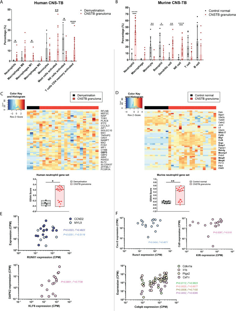
Despite anti-tuberculous treatment (ATT), central nervous system tuberculosis (CNS-TB) still causes permanent neurological deficits and death. To identify prognostic factors, we profiled a prospective cohort of pediatric HIV-negative tuberculous meningitis (TBM) and non-TBM patients. We found significantly increased cerebrospinal fluid (CSF) matrix metalloproteinases (MMPs) and neutrophil extracellular traps (NETs) in TBM patients with neuroradiological abnormalities and poor outcomes. To dissect mechanisms, we used our existing CNS-TB murine model, which shows neutrophil-rich necrotizing pyogranulomas with MMP-9 and NETs colocalizing, as observed in human CNS-TB pathology. Spatial transcriptomic analysis of both human and murine CNS-TB demonstrates a highly-inflamed and neutrophil-rich microenvironment of inflammatory immune responses, extracellular matrix degradation and angiogenesis within CNS-TB granulomas. Murine CNS-TB treated with ATT and MMP inhibitors SB-3CT or doxycycline show significantly suppressed NETs with improved survival. MMP inhibition arms show attenuated inflammation and well-formed blood vessels within granulomas. Adjunctive doxycycline is highly promising to improve CNS-TB outcomes and survival.

Keywords: Central nervous system tuberculosis; Doxycycline; Host-directed therapy; Matrix metalloproteinases; Neutrophil extracellular trap.


Conflict of interest statement

Declaration. Ethics approval and consent to participate: The Domain Specific Review Board from National Healthcare Group Singapore (Reference: 2015/00067) approved the study. Anonymized human adult brain samples were used. All animal procedures were approved by the Institutional Animal Care and Use Committee of National University of Singapore under protocols R15-1068 and R21-0633, in accordance with national guidelines for the care and use of laboratory animals for scientific purposes. Consent for publication: Informed consent was received from all the patients including consent for publication. Competing interests: The authors declare no competing interests.

Figures

Fig. 1
Fig. 1
Up-regulated MMP and NET concentration in human pediatric TBM patients are strongly associated with neuroradiological abnormalities and poor clinical outcomes. A CSF samples from a pediatric cohort of TBM patients and non-TBM patients were analyzed for the expression of MMPs, NETs, and functional gelatinase activity. The CT brain images of TBM patients were assessed for radiological abnormalities together with clinical outcomes. Illustration created using Biorender. B CSF concentrations of MMP-9, MMP-2 and EMMPRIN are upregulated in human pediatric TBM. C CSF concentrations of TIMP-2 and TIMP-4 are downregulated in TBM. In B and C, bars represent median ± IQR with analysis by Mann-Whitney test. Non-TBM, n = 52; TBM, n = 20. D MMP-2 and − 9 neutralization significantly suppress CSF gelatinase activity in TBM patients. Analysis by paired t-test. MMP-2, n = 4; MMP-9, n = 6. E Representative CT brain imaging of human pediatric TBM patients. Left panel shows normal CT brain in a pediatric TBM patient. Right panel shows widespread leptomeningeal enhancement (arrow) and dilated ventricles from hydrocephalus (H) in a pediatric TBM patient. F Increased CSF MMPs significantly associate with leptomeningeal enhancement, ventricular dilatation and poor clinical outcomes in human TBM. Leptomeningeal enhancement, n = 14; ventricular dilatation, n = 14; clinical outcomes n = 17. Bars represent median ± interquartile range (IQR), dots represent individual values. Analysis by Mann-Whitney test. G NETs marker extracellular DNA (n = 52 non-TBM, 20 TBM) and citH3 (n = 6 per group) are increased in human TBM. Bars represent median ± IQR. Analysis by Mann-Whitney test. H CSF CitH3 concentration strongly associates with CSF extracellular DNA concentration and CSF MMP-9 concentrations. TBM patients form a cluster with high NET and MMP-9 concentrations (circled). Analysis by Pearson correlation coefficient. I High CSF extracellular DNA significantly associate with leptomeningeal enhancement, ventricular dilation and poor clinical outcomes in human TBM. Bars represent median ± IQR, dots represent individual values. Statistical analysis was conducted using Mann-Whitney test. * P < 0.05; ** P < 0.01, *** P < 0.001
Fig. 2
Fig. 2
Functional gelatinase activity is increased with H37Rv infection and is associated with MMP-2 and − 9 concentrations in murine CNS-TB. Six- to eight-week-old male C57BL6 Nos2−/− were infected with 105 CFU M. tb H37Rv via intracerebroventricular (i.c.vent). route, control mice received saline (n = 6). Eight weeks post-infection, the brain tissues and homogenates were used for downstream assay. Brain concentrations of (A) gelatinase MMP-2, −9, stromelysin MMP-3, and elastase MMP-12, and (B) TIMP-2 and − 4 in saline control versus H37Rv i.c.vent.-infected Nos2−/− mice. MMP and TIMP concentrations are normalized to total protein concentration. Bars represent median ± IQR, and analysis was conducted using Mann-Whitney test. C Functional gelatinase activity in the brain homogenates of H37Rv i.c.vent.-infected Nos2−/− mice is significantly increased. Bars represent mean ± standard deviation (SD). Analysis by unpaired t-test. D Brain gelatinase activity is associated with MMP-9 concentration, but shows no significant association with MMP-2 in M. tb-infected Nos2−/− mice, analyzed by Spearman’s correlation coefficient. E Brain concentration of NETs marker CitH3 is significantly up-regulated in H37Rv i.c.vent.-infected Nos2−/− mice relative to saline control. Bars represent median ± IQR. Analysis by Mann-Whitney test. F Brain MMP-9 is associated with NETs marker CitH3 concentrations in M. tb-infected Nos2−/− mice. Analysis by Spearman’s correlation coefficient. * P < 0.05; ** P < 0.01. G Representative immunohistochemistry (IHC) stains of granulomas in human CNS-TB (n = 6) and murine CNS-TB (n = 6) show expression of MMP-9 and CitH3 at the necrotic area. Top panel, scale bar = 100 μm; Bottom panel, scale bar = 200 μm
Fig. 3
Fig. 3
Spatial transcriptomic profiling of human and murine CNS-TB shows enrichment of inflammatory and immune genes and pathways. A Target regions of formalin-fixed paraffin-embedded (FFPE) CNS tissue samples from human CNS-TB (n = 6), control human demyelination tissues (n = 4), murine CNS-TB (n = 4) and healthy murine controls (n = 4) were microdissected for LCM RNA-Seq. Illustration created using Biorender. B PCA plot of gene expression libraries demonstrates clear separation of human CNS-TB granulomas and CNS-TB inflammatory from demyelination and adjacent normal tissues. C Hierarchical clustering of top 500 variable genes expressed in human CNS-TB. Red frame highlights gene cluster that is distinctly expressed in human CNS-TB granulomas compared to other tissues. D Volcano plot of up-regulated (log fold-change > 2, FDR < 0.05) and down-regulated (log fold-change < −2, FDR < 0.05) genes in human CNS-TB granulomas compared to adjacent normal tissues. E PCA plot of gene expression libraries showing distinct clustering of murine CNS-TB granulomas and inflammatory tissues from adjacent normal tissues and normal tissue from healthy murine controls. F Hierarchical clustering of top 500 variable genes expressed in murine samples. Red and blue frames highlight gene cluster that are distinctly up-regulated and down-regulated, respectively, in murine CNS-TB granulomas relative to other tissue types. G Volcano plot of up-regulated (log fold-change > 2, FDR < 0.05) and down-regulated (log fold-change < 2, FDR < 0.05) genes in murine CNS-TB granulomas compared to normal tissues from healthy murine controls
Fig. 4
Fig. 4
Key immune cells, neutrophil marker genes and transcription factors driving immunopathology in human and murine CNS-TB granulomas. A Deconvolution analysis by CIBERSORTx LM22 signature reveals that relative to human demyelination tissue, the in silico cell composition in human TB granulomas has higher fractions of neutrophils, macrophages M1, activated mast cells and activated CD4 memory T cells. Bars represent mean ± SD, and analysis was conducted using unpaired t test with Welch’s correction. * P < 0.05; ** P < 0.01; *** P < 0.001; **** P < 0.0001. B In silico deconvolution analysis using ImmuCC signature matrix indicates that relative to healthy murine brain tissues, murine CNS-TB granulomas consist mainly of neutrophils, dendritic cells and NK cells. Bars represent mean ± SD, and analysis was conducted using unpaired t-test with Welch’s correction. C-D Heatmap shows higher expression of neutrophil genes in (C) human CNS-TB granuloma samples compared to demyelination samples and (D) murine CNS-TB granuloma samples compared to control normal samples. Genes in bold font are significantly upregulated with FDR < 0.05. Enrichment of neutrophil gene sets is analyzed by gene set variation analysis (GSVA). Box represents 25th and 75th percentile, line represents median and whiskers denote the extremes. Analysis using Mann-Whitney test. * P < 0.05; ** P < 0.01. E-F Key transcriptional factors, which regulate neutrophil functions, correlate significantly with expression of their neutrophil-associated target genes in human and murine CNS-TB granulomas. Analysis by Spearman’s correlation coefficient
Fig. 5
Fig. 5
Adjunctive MMP inhibition with SB-3CT or doxycycline with standard ATT significantly improves murine CNS-TB survival and reduces immunopathology with decreased expression of NETs and inflammatory mediators. A Six- to eight-week-old male C57BL/6 Nos2−/− mice were infected with 105 CFU M. tb H37Rv via the i.c.vent. route and treated with ATT, ATT + SB-3CT or ATT + Doxycycline from 3 weeks post-infection. (n = 7 to 8 per group). After eight weeks of treatment, the brain homogenates were collected for downstream analyses. Illustration created using Biorender. B Kaplan–Meier curve shows a significant improvement in survival time of M. tb-infected mice treated with MMP inhibitor SB-3CT or doxycycline plus ATT. Log rank test was performed. C Representative hematoxylin–eosin stains of M. tb-infected mouse brain pyogranulomas in VC (infected) and ATT alone groups (top panel), and non-necrotizing granulomas in ATT + SB-3CT and ATT + Doxycycline groups (bottom panel). Scale bar = 100 μm. D Histopathological severity scoring of brain lesions is higher in vehicle control (M. tb-infected) and ATT-alone mice with lower severity scores in adjunctive MMP inhibitor-treated mice. Bars represent median ± IQR. Analysis was conducted using Kruskal-Wallis test with Dunn’s multiple comparisons test. E Granuloma numbers are less in M. tb-infected mice treated with ATT with adjunctive MMP inhibitors. Bars represent median ± IQR, analysis was conducted using Kruskal-Wallis test with Dunn’s multiple comparisons test. F Ziehl–Neelsen staining shows the presence of AFB in the granuloma of murine CNS-TB of different groups. Scale bar = 10 μm. G Smaller number of AFB-containing granulomas are found in mice on ATT and adjunctive MMP inhibitor SB-3CT or doxycycline. Bars represent median ± IQR, analysis was conducted using Kruskal-Wallis test with Dunn’s multiple comparisons test. H CitH3 expression is significantly up-regulated in brain homogenates of mice with liquefactive necrosis. Bars represent median ± IQR. Analysis by Mann-Whitney test. I CitH3 expression is suppressed in CNS-TB mice treated with adjunctive MMP inhibitors SB-3CT or doxycycline. Bars represent median ± IQR. Analysis by Mann-Whitney test. J Decreased CitH3 in the brain homogenates of murine CNS-TB is strongly associated with longer survival days. Analysis was conducted using Spearman correlation coefficient. K Brain homogenate MMP-9 is strongly associated with CitH3, analyzed by Spearman correlation coefficient. L Heatmap shows suppressed median expression of inflammatory mediators in adjunctive MMP inhibitor groups. M Inflammatory mediators TNF-α, IFN-ɣ, CXCL2, CXCL10, CCL2, CCL3, CCL7, CCL11, and CCL19 are inversely correlated with longer survival days (all strong associations with rs < −0.5), analyzed by Spearman correlation coefficient. * P < 0.05; ** P < 0.01
Fig. 6
Fig. 6
Adjunctive doxycycline with ATT suppresses functional gelatinase activity and normalizes blood vasculature in murine CNS-TB granulomas. A Heatmap showing median expression of brain MMP-2, −9, −8, −3, −10, and − 12 downregulated in the MMP inhibitor groups of murine CNS-TB. MMP concentrations are normalized to total protein concentration. B Suppressed murine brain homogenate MMP-3, −8, −9, 10, and − 12 are strongly associated with longer survival, analyzed by Spearman correlation coefficient. C Up-regulated MMP-3, −9, and − 12 are significantly associated with worse histopathological scores, analyzed by Spearman correlation coefficient. D Functional gelatinase activity is suppressed in the brain homogenates of murine CNS-TB which received ATT, ATT + SB-3CT or ATT + Doxycycline. Bars represent mean ± SD, analysis was conducted using one-way analysis of variance (ANOVA) with Dunn’s multiple comparisons test. E Brain homogenate MMP-9 of murine CNS-TB is strongly associated with functional gelatinase activity, analyzed by Spearman correlation coefficient. F The functional gelatinase activity of murine brain homogenates is strongly associated with longer survival, analyzed by Pearson correlation coefficient. G In silico deconvolution analysis using ImmuCC signature matrix was conducted to infer immune cell fractions in murine CNS-TB of different treatment groups. Bars represent mean ± SD, and analysis was conducted using two-way ANOVA with Dunn’s multiple comparisons test. H Enrichment of Gene sets using the transcriptome data from murine CNS-TB treated with different regimes was analyzed by GSVA. Box represents 25th and 75th percentile, line represents median and whiskers denote the extremes. Analysis using unpaired t-tests. * P < 0.05; ** P < 0.01, *** P < 0.001. I Blood vessels were double-stained for endothelial cells with CD31and alpha-smooth muscle actin (α-SMA) for pericytes in the human CNS-TB brain. Inset shows the granuloma rim and center. Vessels with wide lumen are distributed at the rim and mostly poorly-formed microvessels are found at the center. Gray outline indicates granuloma rim. Orange arrow points to the blood vessel. Scale bar = 200 μm. J Compressed vessels and poorly formed microvessels are found in the granuloma periphery of M. tb-infected mice treated with VC or ATT alone. Conversely, vessels with open lumen are present in the granuloma center of M. tb-infected mice received adjunctive SB-3CT or doxycycline. Yellow “c” indicates granuloma center. Scale bar = 200 μm. K CNS-TB mice treated with ATT + Doxycycline have significantly increased total area of blood vessels with pericyte coverage compared to mice that received vehicle control or ATT alone. In each group, four to six tissue sections with the presence of granulomas are assessed in a blinded fashion. Bars represent median ± IQR, analysis was conducted using Kruskal-Wallis test with Dunn’s multiple comparisons test. L VC and ATT alone groups have more vessels located around the periphery, whereas the MMP inhibitor groups have more vessels distributed towards the center of the granuloma. Each point represents the distance of the blood vessel from the granuloma rim. The distance towards the granuloma center is calculated as a negative value. In each group, four to six tissue sections with seven to thirty-eight blood vessels within the maximum range of 300 μm away from the granuloma rim are quantified. Bars represent median ± IQR, analysis was conducted using Kruskal-Wallis test with Dunn’s multiple comparisons test. M Top panel shows representative matrix-assisted laser desorption ionization mass spectrometry imaging (MALDI-MSI) images of murine CNS-TB brain tissues. Rifampicin is evenly distributed in the ATT alone group, in contrast to ATT + SB-3CT and ATT + Doxycycline groups which show higher concentrations within the granuloma. Bottom panel shows respective ZN stains. Red circled area indicates granulomas region with AFB stained. Scale bar = 200 μm

References

    1. Rock RB, Olin M, Baker CA, Molitor TW, Peterson PK. Central nervous system tuberculosis: pathogenesis and clinical aspects. Clin Microbiol Rev. 2008;21(2):243 – 61, table of contents. Epub 2008/04/11. 10.1128/CMR.00042-07. PubMed PMID: 18400795; PubMed Central PMCID: PMCPMC2292571. - PMC - PubMed
    1. Price NM, Farrar J, Tran TT, Nguyen TH, Tran TH, Friedland JS. Identification of a matrix-degrading phenotype in human tuberculosis in vitro and in vivo. J Immunol. 2001;166(6):4223–30. 10.4049/jimmunol.166.6.4223. Epub 2001/03/10. - PubMed
    1. Ong CW, Elkington PT, Friedland JS. Tuberculosis, pulmonary cavitation, and matrix metalloproteinases. Am J Respir Crit Care Med. 2014;190(1):9–18. 10.1164/rccm.201311-2106. Epub 2014/04/10. - PMC - PubMed
    1. Poh XY, Loh FK, Friedland JS, Ong CWM. Neutrophil-Mediated immunopathology and matrix metalloproteinases in central nervous System - Tuberculosis. Front Immunol. 2021;12:788976. 10.3389/fimmu.2021.788976. Epub 2022/02/01. - PMC - PubMed
    1. Kalita J, Misra UK, Nair PP. Predictors of stroke and its significance in the outcome of tuberculous meningitis. J Stroke Cerebrovasc Dis. 2009;18(4):251–8. PubMed PMID: 19560677. - PubMed

MeSH terms

Substances

LinkOut - more resources