. 2025 Oct 11;23(1):660.
doi: 10.1186/s12951-025-03751-x.

Orally administered degradable nanoarmor-assisted probiotics for remodeling the gut microenvironment and treating osteoporosis

Affiliations

Orally administered degradable nanoarmor-assisted probiotics for remodeling the gut microenvironment and treating osteoporosis

Zhiqiang Li et al. J Nanobiotechnology. .

Abstract

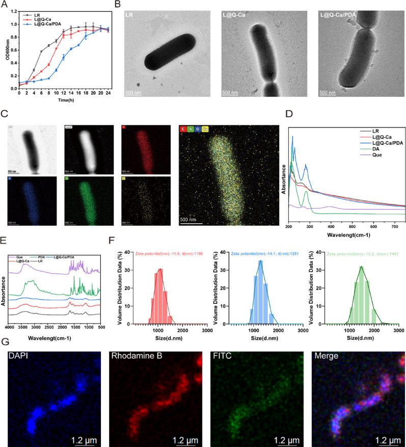
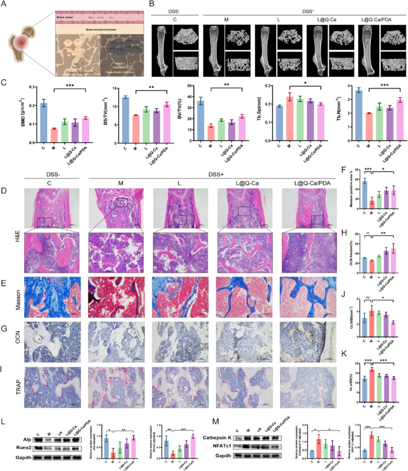
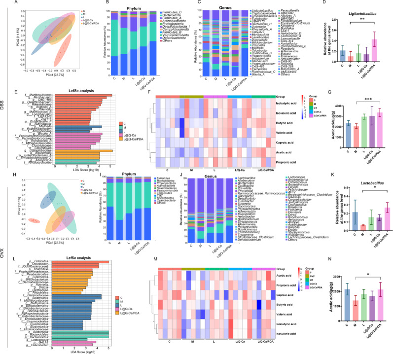
Improving the intestinal microenvironment and regulating the intestinal flora are highly important in the treatment of osteoporosis. Although previous studies have shown that oral probiotics can prevent or reverse bone loss, their survival rate and therapeutic effect are greatly reduced when they pass through the gastrointestinal chemical microenvironment, which limits their clinical application. Therefore, improving their survival rate and therapeutic effect is crucial. To address this issue, we formed a metal‒phenolic network (L@Q-Ca) on the surface of Lactobacillus rhamnosus (LR) by combining quercetin and calcium metal ions to enhance its therapeutic effect. To enable the LR to pass successfully pass the gastrointestinal chemical environment, dopamine was polymerized on the surface of the probiotics, forming a dense protective layer (L@Q-Ca/PDA). Probiotics with the L@Q-Ca/PDA coating significantly outperformed traditional uncoated probiotics in terms of both their survival rate in the gastrointestinal tract and their therapeutic effect on osteoporosis. In the intestinal microenvironment, the composite material can effectively counteract intestinal inflammation, oxidative stress, barrier damage, and microenvironmental disorders. The alleviation of systemic inflammation restores the balance of osteoblast and osteoclast activity. The increased absorption of quercetin and short-chain fatty acids in the intestine can further improve the bone microenvironment. This oral probiotic reinforcement strategy is not only safe, reliable, and efficient, but also potentially amenable to an extremely broad range of applications for the clinical transformation of probiotics in the field of osteoporosis treatment.

Keywords: Bacterial therapeutics; L. rhamnosus; Multifunctional nanocoating; Osteoporosis; Quercetin.


Conflict of interest statement

Declarations. Ethics approval and consent to participate: The experiments complied with the World Medical Association (WMA)’s Statement and China National Standard (GB/T 35892-2018) on animal use in biomedical research. The ethical approval for animal experiments was granted by the Experimental Animal Welfare Ethics Committee of Nanchang University (Approval No. NCULAE-20231124071). Competing interests: All authors declare they have no competing interests.

Figures

Fig. 1
Fig. 1
The key steps and mechanisms of using functionalized probiotics to treat osteoporosis through the “gut-bone” axis
Fig. 2
Fig. 2
Preparation and characterization of L@Q-Ca/PDA. (A) Growth curves of LR, L@Q-Ca, L@Q-Ca/PDA; (B) Representative TEM images of LR, L@Q-Ca, L@Q-Ca/PDA; (C) HAADF-STEM image and corresponding elemental mapping images of C, N, O, and Ca; (D) UV–vis absorption spectra of LR, L@Q-Ca, L@Q-Ca/PDA; (E) Fourier transform infrared spectra of LR, L@Q-Ca, L@Q-Ca/PDA; (F) Zeta potentials and size distributions of the samples measured by DLS; (G) Typical confocal images of L@Q-Ca/PDA. Scale bar: 1.2 μm. The red channel shows rhodamine B-labeled MPNs and the green channel indicates PDA-incorporated coatings
Fig. 3
Fig. 3
Resistance of L@Q-Ca/PDA Against Environmental Assaults and Mucoadhesive Capability. Probiotics were separately exposed to harsh environments as follows: (A) Schematic illustration of the awakening process; (B) SGF (pH 2.0), (C) bile salt (0.3 mg·mL − 1), and (D) SIF (pH 6.8); (E) TEM images of L@Q-Ca/PDA after incubation in SGF for 0 h, SGF for 4 h and SIF for 6 h, respectively; (F) Adhesion of probiotic strains in each group on Caco-2 cells observed by CLSM. Green: Caco-2 stained with fluorescein isothiocyanate (FITC). Red: rhodamine B-labeled probiotics; (G) Representative IVIS images of mice administered with various rhodamine B-labeled probiotic formulations; (H) IVIS images of mouse gut at 120 h post-administration of various rhodamine B-labeled probiotic formulations; (I) Region-of-interest analysis of fluorescence intensity of the mouse GIT at 120 h. Statistical analysis was performed using one-way ANOVA.*P < 0.05, **P < 0.01, ***P < 0.001, ****P < 0.0001
Fig. 4
Fig. 4
Anti-inflammatory and antioxidant effects of L@Q-Ca/PDA in vitro. (A) Schematic illustration of the experimental design; (B) Intracellular ROS levels of H2O2-treated RAW 264.7 cells after different treatments; Antioxidant-related indices were detected, including MDA (C), T-AOC (D), SOD (E), and GSH (F); (G-J) RT-qPCR analysis of IL-10 (G), TGF-β (H), TNF-α (I), and IL-6 (J) mRNA expression in RAW 264.7 cells after different treatments as indicated; (K) iNOS and CD206 fluorescence images of H2O2-treated RAW 264.7 cells after different treatments. Statistical analysis was performed using one-way ANOVA. *P < 0.05, **P < 0.01, ***P < 0.001, ****P < 0.0001
Fig. 5
Fig. 5
Therapeutic efficacy of L@Q-Ca/PDA against DSS-induced intestinal mucositis. (A) Schematic illustration of modeling and treatment; (B) Percentage change in body weight of mice and (C) DAI scores during the experiment; (D) Colon images, and (E) quantified colon lengths of mice; Representative H&E staining (F) and AB-PAS staining (G) images of the colon tissue; (H) Fluorescence images and quantitative analysis (I) of TUNEL staining in colon tissue; (J) Immunofluorescence images and quantitative analysis of the expression of ZO-1 (K) and Occludin (L) in the colon; Expression of the proinflammatory cytokines TNF-α (M) ,IL-1β (N) and IL-6 (O) in the blood of mice. Statistical analysis was performed using one-way ANOVA. *P < 0.05, **P < 0.01, ***P < 0.001, ****P < 0.0001.
Fig. 6
Fig. 6
Therapeutic efficacy of L@Q-Ca/PDA in osteoporosis in model mice with colitis-induced osteoporosis. (A) Schematic illustration of systemic inflammation relief in osteoporosis; (B) Micro-CT images of the distal femur in different groups; (C) BMD, BV/TV, BS/TV, Tb.N, and Tb.Sp structural parameters in different groups of DSS-induced colitis mice; Representative H&E staining (D), Masson staining (E, F), OCN staining (G, H), and TRAP staining (I-K) images of bone tissue and their quantitative results; (L) Western blotting analysis of osteogenic factors (RUNX2 and ALP) with protein levels and quantitative results; (M) Western blotting analysis of osteoclast factors (Cathepsin K and NFATc1) with protein levels and quantitative results; Statistical analysis was performed using one-way ANOVA. *P < 0.05, **P < 0.01, ***P < 0.001, ****P < 0.0001
Fig. 7
Fig. 7
Therapeutic efficacy of L@Q-Ca/PDA in model mice with OVX Osteoporosis. (A) Schematic illustration of modeling and treatment; (B) Wet weight of the uterus and (C) body weight of mice during the experiment; (D) Micro-CT images of the distal femur in different groups and (E) their quantitative results; Representative H&E staining (F), Masson staining (G, H), OCN staining (I, J), and TRAP staining (K, L, M) images of bone tissue and their relative quantitative results; Representative H&E staining (N), AB-PAS staining (O), TUNEL staining (P, Q), and ZO-1/Occludin staining (R, S, T) images of colon tissue and their relative quantitative results; (U) Western blotting analysis of osteogenic factors (RUNX2 and ALP) with protein levels and quantitative results; (V) Western blotting analysis of osteoclast factors (Cathepsin K and NFATc1) with protein levels and quantitative results; Statistical analysis was performed using one-way ANOVA. *P < 0.05, **P < 0.01, ***P < 0.001, ****P < 0.0001
Fig. 8
Fig. 8
Regulation of L@Q-Ca/PDA on gut microbiome homeostasis in mouse models of colitis-induced osteoporosis and OVX-induced osteoporosis. (A, H) PCoA score plots illustrating the β-diversity of the gut microbiome; (B, I) Relative abundances of gut commensal microbes at the phyla levels after different treatments; (C, J) Relative abundances of gut microbiota at the genus level; (D, K) Relative contents of Lactobacillus in fecal samples; (E, L) Distribution histograms based on linear discriminant analysis (LDA). LDA scores higher than 2.0 indicate taxa that are significantly more abundant in the corresponding group than in other groups; (F, M) Heatmap illustrations of short-chain fatty acids; (G, N) Relative contents of acetic acid in fecal samples; Statistical analysis was performed using Student’s t-test. * P < 0.05, **P < 0.01, ***P < 0.001, ****P < 0.0001

References

    1. Reid IR. A broader strategy for osteoporosis interventions. Nat Rev Endocrinol [Internet]. 2020 [cited 2024 Nov 28];16:333–9. Available from: https://www.nature.com/articles/s41574-020-0339-7 - PubMed
    1. Yang X, Fan Y, Liang J, Cao R, Zhang B, Li J, et al. Polyaptamer-driven crystallization of alendronate for synergistic osteoporosis treatment through osteoclastic inhibition and osteogenic promotion. ACS Nano. 2024;18:22431–43. 10.1021/acsnano.4c07265. - PubMed
    1. Clynes MA, Harvey NC, Curtis EM, Fuggle NR, Dennison EM, Cooper C. The epidemiology of osteoporosis. British Medical Bulletin [Internet]. 2020 [cited 2024 Nov 29];ldaa005. Available from: https://academic.oup.com/bmb/advance-article/doi/10.1093/bmb/ldaa005/5817480 - PMC - PubMed
    1. Reid IR. A broader strategy for osteoporosis interventions. Nat Rev Endocrinol [Internet]. 2020 [cited 2024 Nov 29];16:333–9. Available from: https://www.nature.com/articles/s41574-020-0339-7 - PubMed
    1. Amigues C, Fresse A, Roux C, Gauthier S, Vieillard MH, Drici MD. OP0240 ZOLEDRONATE-RELATED OSTEONECROSIS OF THE JAW IN OSTEOPOROSIS: INCIDENCE, RISK FACTORS AND COMPARISON TO ORAL BISPHOSPHONATES. Ann Rheum Dis. 2022;81:158–158. 10.1136/annrheumdis-2022-eular.2876.

LinkOut - more resources