. 2025 Sep 26:16:1618677.
doi: 10.3389/fimmu.2025.1618677. eCollection 2025.

HuR ablation destabilizes Foxp3 mRNA and impairs regulatory T cell function, contributing to an autoimmune phenotype

Affiliations

HuR ablation destabilizes Foxp3 mRNA and impairs regulatory T cell function, contributing to an autoimmune phenotype

Fatemeh Fattahi et al. Front Immunol. .

Abstract

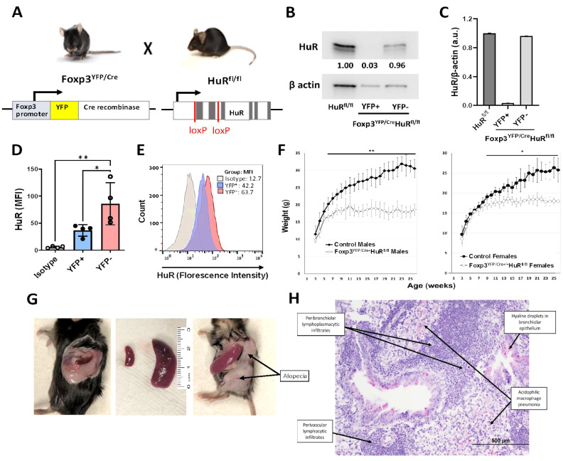
Introduction: The RNA-binding protein HuR (Elavl1), a key post-transcriptional regulator, plays a critical role in T cell activation and function by stabilizing target mRNAs. To investigate the role of HuR in regulatory T cell (Treg) function, we generated the Foxp3YFP/Cre HuRfl/fl mouse model.

Methods: In this model, homozygous females and hemizygous males for Foxp3 developed a scurfy-like phenotype displaying autoimmune features, including failure to thrive, splenomegaly, hair loss, tail stippling, and widespread multi-organ immune cell infiltration. Molecular analysis included direct interaction studies between HuR and Foxp3 mRNA to assess mRNA stability, RNA sequencing of YFP⁺ Tregs, Protein-Protein Interaction (PPI) analysis, qPCR, and Treg functional assays.

Results: To our knowledge, this is the first study demonstrating that HuR directly binds and stabilizes Foxp3 mRNA in Tregs, using a novel Treg-specific HuR-deficient mouse model, with implications for autoimmune regulation. Foxp3 mRNA stability and expression were significantly reduced in Tregs from these HuR KO mice, despite higher frequencies of YFP⁺ Tregs. RNA sequencing revealed significant dysregulation of several pathways, including the T helper differentiation pathway, in which Foxp3 played a central role. PPI analysis showed a direct link between Foxp3 and Rorc (encoding RORγt), connecting Foxp3 to the T cell differentiation pathway via IL-23R. Our qPCR analysis supported these findings. Functional assays demonstrated a reduction in the suppressive capacity of HuR-deficient Tregs.

Conclusion: These findings together suggest that ablation of HuR in Tregs disrupts Foxp3 expression and Treg function, likely through dysregulation of T cell differentiation pathways involving RORγt. This potentially contributes to a disrupted Treg-Th17 axis and autoimmune dysfunction. These data suggest that HuR-mediated post-transcriptional regulation contributes to maintaining Foxp3 expression and immune homeostasis, although compensatory mechanisms such as increased IL-10 expression may also be involved.

Keywords: Foxp3 stability; HuR (ELAVL1); IPEX syndrome; RNA-binding proteins (RBPs); RORγt (Rorc); post-transcriptional regulation; regulatory T cells (Tregs); systemic autoimmunity.


Conflict of interest statement

The authors declare that the research was conducted in the absence of any commercial or financial relationships that could be construed as a potential conflict of interest.

Figures

Figure 1
Figure 1
Treg-specific ablation of HuR in Foxp3 YFP/Cre HuR fl/fl mice results in severe failure to thrive (FTT) and systemic spontaneous autoimmunity. (A) Schematic illustrating conditional deletion of HuR in Tregs in the Foxp3 YFP/Cre , HuR fl/fl (HuR-KO Tregs) mouse model. (B) Western blot of HuR protein in sorted YFP+ (HuR-null) versus YFP Treg populations showing near-complete HuR deletion in YFP+ cells, with β-actin as loading control. (C) Quantification of HuR/β-actin protein levels from western blots, showing HuR reduction in YFP+ Tregs compared to controls. (D) Bar graph of HuR mean fluorescence intensity (MFI) in YFP+ versus YFP Tregs and isotype control, showing significant HuR reduction in YFP+ Tregs (mean ± SD, p < 0.05 by unpaired t-test, n = 4). (E) Flow cytometry histogram representative showing HuR expression profiles in YFP+, YFP, and isotype control populations. (F) Body weight monitored from 3–26 weeks in male and female Foxp3 YFP/Cre , HuR fl/fl (open symbols) versus controls (solid symbols). Both male and female HuR-deficient mice exhibited severe growth impairment (males plateaued at 18.5 ± 1.2 g vs. 31.5 ± 1.6 g in controls, p < 0.01; females plateaued at 18.3 ± 1.0 g vs. 26.5 ± 2.4 g in controls, p < 0.05). Two-way repeated measures ANOVA showed significant genotype effect. (G) Clinical phenotype in Foxp3 YFP/Cre , HuR fl/fl mice including splenomegaly, alopecia, and tail stippling. (H) Representative H&E lung section from Foxp3 YFP/Cre , HuR fl/fl mice demonstrating perivascular and peribronchiolar lymphoplasmacytic infiltrates, acidophilic macrophage pneumonia, and hyaline droplets in bronchiolar epithelium. Representative data are shown from at least 8 mice for panels F-H. The age of the mice at the time of analysis was 6–7 months. *p<0.05, **p<0.01.
Figure 2
Figure 2
HuR directly interacts with Foxp3 mRNA and controls its steady-state levels and mRNA stability. (A) Foxp3 mRNA stability was evaluated in splenic Tregs (CD4+CD25+) isolated by magnetic column separation from WT mice treated with KH-3 or sham control (KH3-B) for 2 hours, followed by actinomycin (D) HuR inhibition reduced Foxp3 mRNA half-life (1.9 hours with KH-3) compared to control (4.3 hours). Data represents seven WT mice. (B) Foxp3 mRNA stability was assessed in column-isolated splenic Tregs from WT and Foxp3 YFP/Cre HuR fl/fl (HuR-KO Tregs) mice. HuR-KO Tregs showed a shorter Foxp3 mRNA half-life (1.9 hours) compared to WT (4.2 hours). Data represents seven WT and four HuR-KO mice. (C) Steady-state Foxp3 mRNA levels were measured in splenic and thymic Tregs. Foxp3 mRNA was significantly lower in HuR-KO Tregs compared to WT control, while CD4+CD25 cells showed minimal expression with no significant differences. Data represent three HuR-KO and three WT mice. (D) RNA immunoprecipitation (RIP) from WT splenic Tregs showed Foxp3 mRNA enrichment in HuR pull-downs compared to IgG1 control, with β-actin as a positive control. Each point represents pooled cells from four WT mice. A total of 12 mice were used across three independent experiments. **p<0.01, ****p<0.0001.
Figure 3
Figure 3
Gene profiling of RNA-seq and PPI analysis reveals direct interaction between Rorc and Foxp3 in HuR-KO Tregs in Foxp3 YFP/Cre HuR fl/fl Tregs. (A) Volcano plot visualization of RNA-Seq results from sorted YFP+ (HuR-KO Tregs) isolated from the spleen of Foxp3 YFP/Cre HuR fl/fl mice. The plot shows the expression levels of 2220 statistically significant genes in YFP+ (HuR-KO Tregs), with differentially expressed genes highlighted (upregulated in red, downregulated in blue). FOXP3, Rorc, and IL-23R, were identified and labeled. (B) Protein-protein interaction (PPI) network analysis using Cytoscape identified significant gene interactions. Among the differentially expressed genes in YFP+ (HuR-KO Tregs), Rorc (RORγt) was found to directly interact with FOXP3, as shown in the enlarged region (red dashed box). n = 6 mice.
Figure 4
Figure 4
Altered mRNA expression of selected genes involved in T helper differentiation and Treg function in Foxp3 YFP/Cre HuR fl/fl Tregs compared to WT control Tregs. (A-F) mRNA expression of key genes involved in T helper differentiation (based on RNA-seq IPA analysis) and Treg function including Rorc, Il23r, Ctla4, Il2r, Il10 and Il17a was measured by qPCR in CD4+CD25+ Tregs isolated from the spleens of HuR-KO and WT control mice, following 4 days of activation with anti-CD3 and anti-CD28. Data were analyzed using unpaired Student’s t-test. Results are representative of four independent HuR-KO mice and a total of six corresponding WT controls, from cohorts distinct from those used in RNA-seq analysis.
Figure 5
Figure 5
Impaired suppressive function of HuR-KO Tregs (Foxp3 YFP/Cre HuR fl/fl ) on Teffector cell proliferation. The in vitro suppressive ability of Tregs in regulating Teffector cell (Teff) proliferation was evaluated by flow cytometry. Teff cells were labeled with a proliferation dye prior to co-culture with isolated Tregs (CD4+CD25+) from control HuR fl/fl WT (blue bars) and Foxp3 YFP/Cre HuR fl/fl (red bars) mice at varying Treg: Teff ratios, as indicated on the x-axis. (A) Percentage suppression of Teff cell proliferation by CD4+CD25+ Tregs across different Treg: Teff ratios. HuR-KO Tregs showed a significant, progressive decline in suppressive capacity beginning at the 1:64 ratio, whereas WT Tregs maintained stable suppression even at lower Treg frequencies. (B) Representative flow cytometry histograms of Teff proliferation patterns (gated on live cells), demonstrating reduced suppression by HuR-KO Tregs compared to WT Tregs. Overall, HuR-KO Tregs exhibited significantly reduced suppression of Teff cell proliferation (p < 0.0001). Data are from five mice per group, each run-in duplicate. Data were analyzed using two-way ANOVA followed by Šídák’s multiple comparisons test. *p<0.05; **p<0.01.

References

    1. Sakaguchi S, Vignali DA, Rudensky AY, Niec RE, Waldmann H. The plasticity and stability of regulatory T cells. Nat Rev Immunol. (2013) 13:461–7. doi: 10.1038/nri3464, PMID: - DOI - PubMed
    1. Fontenot JD, Gavin MA, Rudensky AY. Foxp3 programs the development and function of CD4+ss+ regulatory T cells. Nat Immunol. (2003) 4:330–6. doi: 10.1038/ni904, PMID: - DOI - PubMed
    1. Hill JA, Feuerer M, Tash K, Haxhinasto S, Perez J, Melamed R, et al. Foxp3 transcription-factor-dependent and -independent regulation of the regulatory T cell transcriptional signature. Immunity. (2007) 27:786–800. doi: 10.1016/j.immuni.2007.09.010, PMID: - DOI - PubMed
    1. Hori S, Nomura T, Sakaguchi S. Control of regulatory T cell development by the transcription factor Foxp3. Science. (2003) 299:1057–61. doi: 10.1126/science.1079490, PMID: - DOI - PubMed
    1. Clark LB, Appleby MW, Brunkow ME, Wilkinson JE, Ziegler SF, Ramsdell F. Cellular and molecular characterization of the scurfy mouse mutant. J Immunol. (1999) 162:2546–54. doi: 10.4049/jimmunol.162.5.2546, PMID: - DOI - PubMed

MeSH terms