. 2025 Oct 14;44(1):289.
doi: 10.1186/s13046-025-03548-8.

FASN inhibits ferroptosis in breast cancer via USP5 palmitoylation-dependent regulation of GPX4 deubiquitination

Affiliations

FASN inhibits ferroptosis in breast cancer via USP5 palmitoylation-dependent regulation of GPX4 deubiquitination

Zhiwen Qian et al. J Exp Clin Cancer Res. .

Abstract

Increasing studies have reported that dysregulated lipid metabolism is an independent risk factor for breast cancer (BC); it would be, therefore, enlightening to investigate the relationship between metabolic reprogramming and the tumor microenvironment in the future. Ferroptosis, a novel form of programmed cell death, is characterized by glutathione (GSH) depletion and inactivation of glutathione peroxidase 4 (GPX4), the central regulator of the antioxidant system. While the close association between fatty acid metabolism and ferroptosis has been studied in various diseases, the interplay between the key fatty acid metabolic enzyme fatty acid synthase (FASN) and ferroptosis in BC remains unexplored. At the beginning of the current study, we demonstrated that FASN expression positively correlates with an immune-cold tumor microenvironment in BC. Subsequent findings revealed that FASN knockdown promotes GPX4 degradation-induced ferroptosis, thereby enhancing the efficacy of anti-programmed cell death protein 1 (PD-1) immunotherapy. Co-immunoprecipitation coupled with mass spectrometry (IP/MS) and co-IP experiments demonstrated that ubiquitin specific protease 5 (USP5) stabilizes GPX4 by binding to and deubiquitinating it. Furthermore, knockdown of FASN inhibited the palmitoylation of USP5, reducing its interaction with GPX4 and consequently increasing GPX4 ubiquitination and degradation. Our results demonstrate that FASN suppresses ferroptosis in BC by stabilizing GPX4 via USP5-mediated mechanisms, highlighting FASN inhibition as a potential therapeutic approach to enhance immunotherapy response.

Keywords: Breast cancer; FASN; Ferroptosis; Palmitoylation; USP5.


Conflict of interest statement

Declarations. Ethics approval and consent to participate: This study involves human participants and ethical approval for the study of TMAs was granted by the Outdo Biotech Clinical Research Ethics Committee. Ethical approval for the single-cell sequencing data was granted by Clinical Research Ethics Committees at Wuxi Maternal and Child Health Care Hospital (Ethics No. 2024–01-1024–31). Participants gave informed consent to participate in the study before taking part. All animal experiments were approved by the Institutional Animal Care and Use Committee (IACUC) of Jiangnan University (JN. No 20250228b0720731[069]). Consent for publication: Not applicable. Competing interests: The authors declare no competing interests.

Figures

Fig. 1
Fig. 1
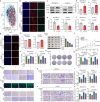
FASN expression patterns and metabolic associations in breast cancer. a, b Association between FASN and fatty acid metabolism pathways including biosynthesis of unsaturated fatty acids (a) and fatty acid summary (b). c scRNA-seq data (n = 19,600 cells from 5 patients) analysis of FASN expression distribution across cellular populations. d Binary classification of cells based on FASN expression (> 0). Bar plots compare FASN + proportions between tumor cells and non-tumor cells. e Multiplex immunofluorescence (mIF) of FASN (magenta) with DAPI (blue). Right: Quantification of FASN + positive area fraction. f, g Representative immunohistochemistry (IHC) and mIF staining of FASN in tumor cells and stromal cells. H&E staining (g, up) serves as spatial reference for mIF analysis. h, i Association between FASN expression and clinicopathological characteristics in TCGA-BRCA cohort. Scale bars: 50 μm (e- g). Groups were compared by Fisher's exact test (d), two-tailed unpaired t-test (e, right) or Wilcoxon rank-sum test (i). P < 0.05 was considered to be statistically significant. IHC images of FASN in tumor and stromal cells were obtained from the Human Protein Atlas (HPA) database (https://www.proteinatlas.org)
Fig. 2
Fig. 2
FASN correlates with tumor immunity in breast cancer. a-c ESTIMATE algorithm-derived tumor microenvironment characteristics in TCGA-BRCA: tumor purity (a), immune score (b), and T-cell inflammation score (c). d Differential expression of immunomodulatory genes between FASN-high and FASN-low groups (median cutoff). e Heatmap of immune cell signature gene expression in FASN-high vs. FASN-low groups (median cutoff). f Correlation between FASN expression and immune cell abundances. g-i Scatter plots showing correlations between FASN expression and CD8+ T-cell abundance predicted by EPIC (g), MCP-counter (h), and TISIDB (i). j, k Representative multiplex immunofluorescence (mIF) images demonstrating expression of FASN and CD8 in tumor cells (j) with Pearson correlation analysis (k). l FASN expression in immunotherapy-treated cohort (CR: complete response; NR: no response). Scale bars: 50 μm (j). Groups were compared by Wilcoxon rank-sum test (d, l). P < 0.05 was considered to be statistically significant
Fig. 3
Fig. 3
FASN modulates breast cancer malignancy by regulating tumor cell proliferation. a Proliferation score analysis of FASN + and FASN − tumor cells in scRNA-seq data (n = 19,600 cells from 5 patients). Proliferation scores were computed by applying the PercentageFeatureSet function in the Seurat package to quantify cell cycle-related gene expression signatures (see Methods). b, c Validation of FASN knockdown efficiency. qRT-PCR analysis of FASN mRNA levels in siRNA-transfected cells (normalized to GAPDH) (b) and western blot analysis of FASN protein expression (β-actin served as loading control) (c). d Apoptosis detection by TUNEL assay in FASN-knockdown cells (48 h post-transfection). Nuclei were stained with DAPI (blue); TUNEL-positive cells (red). e, f Functional consequences of FASN knockdown. e, Clonogenic survival assay (10-day culture). f, Cell viability measured by CCK-8 assay (48 h post-transfection). g Western blot analysis of apoptosis-related proteins (Bax and bcl-2) in FASN-knockdown MDA-MB-231 and SK-BR-3 cells. Quantification of protein levels (normalized to β-actin) is shown below the blots. h, i Effects of FASN knockdown on migration (h) and invasion (i). Representative images (left) and quantifications (right) of Transwell migration (top) and invasion (bottom) assays. Scale bars: 50 μm (h, i). Experiments were performed three times independently (bi). Data were presented as mean ± SD (bf). Groups were compared by two-tailed unpaired t-test (f, right of e, gi) or one-way ANOVA followed by Fisher’s LSD test (b). P < 0.05 was considered to be statistically significant
Fig. 4
Fig. 4
FASN modulates ferroptosis and enhances anti-PD-1 efficacy in breast cancer. a Transcriptomic profiling of breast cancer tumors from the TCGA-BRCA cohort (n = 1,069 patients). Patients were stratified into FASN-high and FASN-low groups based on median FASN expression. t-SNE analysis revealed ferroptosis-related transcriptional patterns between groups. b Lipid peroxidation detection in FASN-knockdown MDA-MB-231 and SK-BR-3 cells using C11-BODIPY 581/591 probe. Representative fluorescence images show oxidized lipid (green) and non-oxidized lipid (red). c Western blot analysis of GPX4 and COX-2 expression in MDA-MB-231 and SK-BR-3 cells following FASN knockdown. d-f Impact of FASN knockdown on oxidative stress and ferroptosis in MDA-MB-231 and SK-BR-3 cells. Cells were treated as in (b), then relative MDA (d), GSH (e), and SOD (f) content were detected by their corresponding kits. g, h FASN knockdown elevates intracellular ferrous iron (Fe2⁺) levels in MDA-MB-231 and SK-BR-3 cells, as quantified by FerroOrange fluorescence (g, 5 μM probe) and ferrozine assay (h). i-m Therapeutic efficacy of orlistat combined with anti-PD-L1 in BALB/c mice bearing 4T1 mammary tumors. Representative tumor images (i, left, n = 5) and comparative tumor weight analysis (i, right) across treatment groups. Tumor growth curves monitored over time (j). Immunohistochemical (IHC) staining of GPX4 and COX-2 in tumor tissues (k). 4-HNE levels detected by immunofluorescence (l, green). IHC analysis of PD-L1 and CD8 expression (m). n-t Effects of FASN knockdown combined with ferrostatin-1 (Fer-1, 5 μM) on malignant behaviors of breast cancer cells, including cell viability (n), clonogenic survival (o, p), migration (q, r) and invasion (s, t). Scale bars: 50 μm (b, g, k-m, q, s). Experiments were performed three times independently (bt). Data were presented as mean ± SD (df, h, i, n, p, r, t). Groups were compared by two-tailed unpaired t-test (df, h) or one-way ANOVA followed by Fisher’s LSD test (i, n, p, r, t). P < 0.05 was considered to be statistically significant
Fig. 5
Fig. 5
FASN knockdown inhibits USP5-mediated GPX4 deubiquitination. a qRT-PCR analysis of GPX4 mRNA in FASN-knockdown MDA-MB-231 and SK-BR-3 cells (normalized to GAPDH). b, c WB and quantification of GPX4 protein degradation in CHX-treated cells (10 μM, indicated times). d, e WB analysis of GPX4 in orlistat-treated cells with MG132 (d, 10 μM, 6 h) or chloroquine (e, CQ, 10 μM, 6 h). f, g Schematic of GPX4 interactor screening by IP-MS (f) and candidate protein list (g). h Co-IP of USP5 with anti-GPX4 antibody in breast cancer cells (IgG control). i, j WB and quantification of GPX4 in USP5-knockdown cells. k IP-WB analysis in siUSP5-expressing cells. l Ubiquitination assays in MG132-treated (10 μM, 6 h) cells transfected with siUSP5. m, n Multiplex immunofluorescence (mIF) images demonstrating expression of USP5 and GPX4 in tumor cells (m) with Pearson correlation analysis (n). o Endogenous GPX4 (red) and USP5 (green) co-localization by immunofluorescence (DAPI: blue;). p Representative mIF images demonstrating expression of USP5 and CD8 in tumor cells. Scale bars: 10 μm(o) and 50 μm (p). Experiments were performed three times independently (al, o). Data were presented as mean ± SD (a, i, l, o). Groups were compared by two-tailed unpaired t-test (a, j). P < 0.05 was considered to be statistically significant
Fig. 6
Fig. 6
USP5 stabilizes GPX4 by deubiquitination in a FASN-dependent manner. a Schematic of USP5 and its truncation mutants. b Domain architecture of GPX4 and truncation mutants. c Co-IP in MDA-MB-231/SK-BR-3 cells co-transfected with HA-GPX4 (or mutants) and Flag-USP5 using anti-Flag antibody. d Reverse Co-IP with anti-HA antibody in cells expressing Flag-USP5 (or mutants) and HA-GPX4. e Cartoon model of GPX4-USP5 interaction. f Local interaction interface from molecular docking. g, h WB and quantification of USP5/GPX4 in FASN-KD cells. i Docking result (Surface + Cartoon mode). j IP-WB analysis in siFASN-expressing cells. k Ubiquitination assay in siFASN cells treated with MG132 (10 μM, 6 h). l GPX4 protein levels upon USP5 overexpression and FASN knockdown. m IF staining of endogenous GPX4/USP5 in FASN-KD cells. n Colocalization of ectopically expressed GPX4 and USP5. Scale bars: 10 μm(m) and 50 μm (n). Experiments were performed three times independently (c, d, g, j, k). Data were presented as mean ± SD (g). Groups were compared by two-tailed unpaired t-test (g). P < 0.05 was considered to be statistically significant
Fig. 7
Fig. 7
Palmitoylation of USP5 regulates GPX4 stability to suppress ferroptosis. a Breast cancer cells transfected with siRNA for 48 h were treated with MG132 (10 μM) for 6 h. USP5 palmitoylation was detected by acyl-biotin exchange (ABE) assay following immunoprecipitation (IP) with anti-USP5 antibody (hydroxylamine (HAM) removed palmitic acid (PA), free thiols labeled with BMCC-biotin, visualized by streptavidin-HRP). b Cells pretreated with 2-bromopalmitate (2-BP, 10 μM) for 48 h followed by MG132 (10 μM) for 6 h were subjected to ABE assay for USP5 palmitoylation (methods as in a). c GPX4 protein levels were analyzed by anti-GPX4 IP after 2-BP (10 μM) treatment for 48 h. d GPX4 ubiquitination was examined in 2-BP-pretreated (as in b) cells following MG132 treatment. e Immunofluorescence (IF) staining of USP5 (green) and GPX4 (red) in 2-BP-treated cells (nuclei: DAPI, blue). f Prediction of USP5 palmitoylation sites by GPS/CSS algorithms. g Schematic of wild-type (WT) USP5 and palmitoylation-deficient mutant (mut USP5). h ABE assay detecting palmitoylation of Flag-tagged WT/mut USP5 after MG132 treatment (IP with anti-Flag, methods as in a). i WB and quantification of Flag-GPX4 in WT/mut USP5-transfected cells. j GPX4 ubiquitination assay in WT/mut USP5-expressing cells treated with MG132. k IF staining of Flag (green) and GPX4 (red) in WT/mut USP5-transfected cells (nuclei: DAPI, blue). l Co-IP of GPX4 in USP5-knockdown cells rescued with WT/mut USP5. m GPX4 ubiquitination assay in rescue experiments (methods as in j). n, o Molecular docking models of GPX4-mut USP5 interaction (n: Surface + Cartoon representation; o: Key interfacial residues). p Working model: FASN promotes USP5 palmitoylation to enhance GPX4 binding, inhibiting GPX4 ubiquitination and subsequent degradation, thereby suppressing ferroptosis. Scale bars: 10 μm (e, k). Experiments were performed three times independently (a-d, h-m). Data were presented as mean ± SD (i). Groups were compared by two-tailed unpaired t-test (i). P < 0.05 was considered to be statistically significant

References

    1. Bray F, et al. Global cancer statistics 2022: GLOBOCAN estimates of incidence and mortality worldwide for 36 cancers in 185 countries. CA Cancer J Clin. 2024;74:229–63. - PubMed
    1. Xu Y, Gong M, Wang Y, Yang Y, Liu S, Zeng Q. Global trends and forecasts of breast cancer incidence and deaths. Sci Data. 2023;10:334. - PMC - PubMed
    1. Łukasiewicz S, Czeczelewski M, Forma A, Baj J, Sitarz R, Stanisławek A. Breast cancer-epidemiology, risk factors, classification, prognostic markers, and current treatment strategies-an updated review. Cancers (Basel). 2021. 10.3390/cancers13174287. - PMC - PubMed
    1. Loibl S, et al. Early breast cancer: ESMO clinical practice guideline for diagnosis, treatment and follow-up. Ann Oncol. 2024;35:159–82. - PubMed
    1. Gradishar WJ, et al. Breast cancer, version 3.2022, NCCN clinical practice guidelines in oncology. J Natl Compr Canc Netw. 2022;20:691–722. - PubMed

Substances