. 2025 Oct 14;16(1):75.
doi: 10.1186/s13293-025-00760-1.

Cell line-specific estrogen responses uncover functional sex differences in murine macrophages

Affiliations

Cell line-specific estrogen responses uncover functional sex differences in murine macrophages

Alison M Veintimilla et al. Biol Sex Differ. .

Abstract

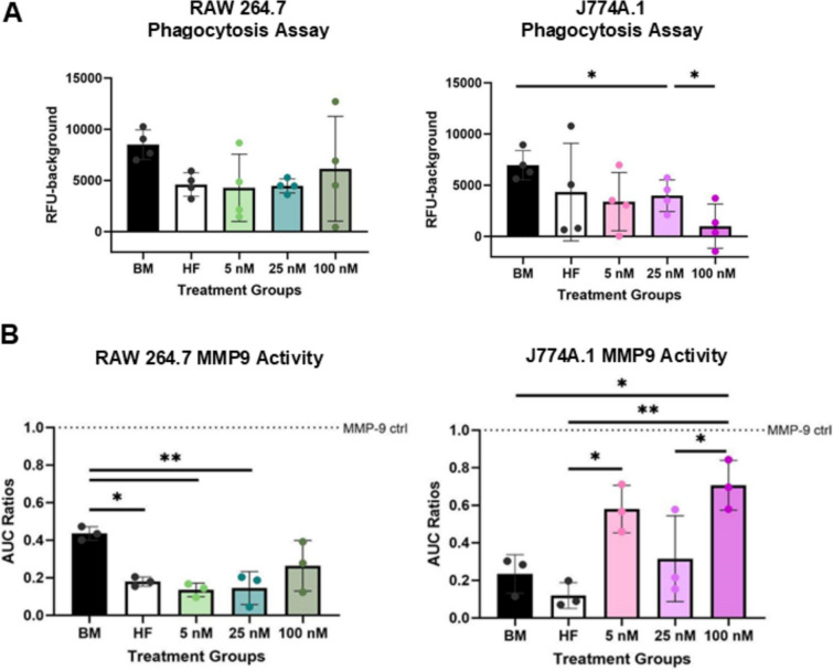
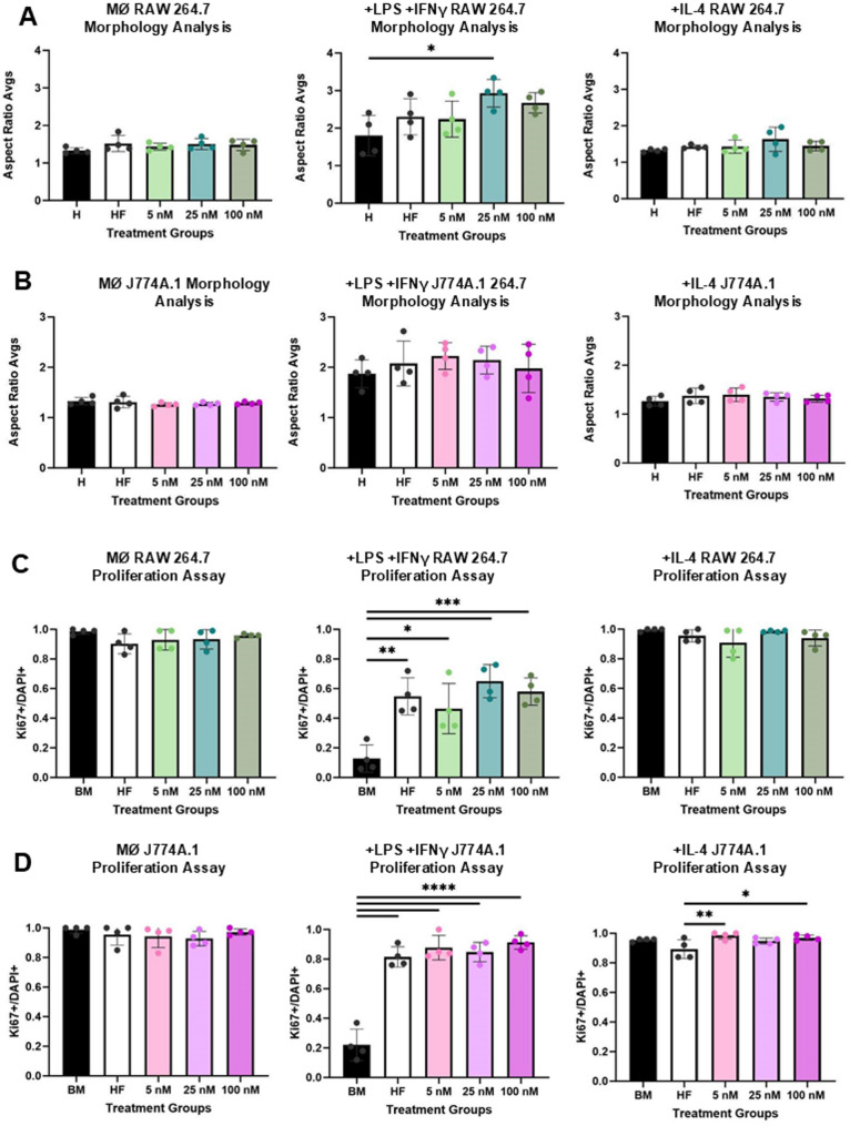
Background: RAW 264.7 (male-derived) and J774A.1 (female-derived) cell lines are widely used in immunology research and are considered preferred models for studying signaling pathways, yet their responses to gonadal hormones remain poorly understood. Gonadal hormones, particularly estrogen, shape immune cell function and contribute to sex differences in disease outcomes, with macrophages playing a central role through their expression of intracellular estrogen receptors (ERs). Herein, we investigated ER expression and functional responses to 17β-estradiol (E2) in male-derived RAW 264.7 and female-derived J774A.1 macrophages, in 2D culture. Additionally, we looked at sex-matched and mismatched media conditions in a 3D hydrogel system. Our results reveal distinct phenotypic and functional differences between the cell lines, emphasizing the need for sex-aware approaches in immunological research and model design.

Methods: RAW 264.7 and J774A.1 macrophages were cultured in basal media for 24 hours, then treated with varying concentrations of 17β-estradiol (5, 25, 100 nM), as well as hormone-free and control media. Post-treatment analyses included viability, estrogen receptor expression, phenotype skewing, matrix metalloprotease 9 (MMP9) activity, and phagocytosis. These macrophages were also used to condition sex-specific media environments and were encapsulated in a hydrogel network containing adhesive and cleavable sites. Encapsulated cells were then exposed to sex-matched or sex-mismatched conditioned media, and proliferation and MMP9 activity were assessed.

Results: Our results revealed distinct differences in estrogen receptor gene and protein expression, as well as in core macrophage functions such as proliferation, inflammation, matrix remodeling, and phenotype skewing. Additionally, the sex-derivation of the surrounding molecular environment affected macrophage behavior in a 3D hydrogel system. Female-derived macrophages were more sensitive in terms of proliferation to sex-mismatched environments, while male-derived macrophages exhibited altered enzyme activity when exposed to female-conditioned media.

Conclusions: These findings underscore the importance of accounting for both the origin of immune cells as well as the hormonal and environmental context in which they are studied. Without these considerations, experimental models risk missing critical biological differences that shape immune responses and disease outcomes.

Keywords: Estrogen; Gonadal hormones; Macrophage function.

Plain language summary

Males and females often experience different symptoms, risks, and outcomes when it comes to certain diseases and health conditions. One reason for this may be that male and female immune cells behave differently, especially in response to hormones like estrogen. In this study, we looked at two commonly used types of mouse immune cells—one originally from a male and one from a female—to see how they respond to estrogen.We found that male and female cells do not respond to estrogen in the same way. They showed different levels of activity, growth, and behavior depending on both the hormone exposure and the sex origin of the environment they were in. We also placed the cells in a gel that mimics tissue and exposed them to sex-specific environments and saw clear differences in how male and female cells responded. For example, female-derived cells were more sensitive in their ability to grow when placed in a “male” environment, while male-derived cells changed their behavior when exposed to signals from a “female” environment.Our findings show that both the origin of immune cells and the environment they are placed in can strongly influence how they behave. This means that to better understand immune responses and develop more effective treatments, scientists need to consider sex as a key factor in their research models. Ignoring these differences could lead to incomplete or misleading results.


Conflict of interest statement

Declarations. Ethics approval: Not applicable. Consent for publication: Not applicable. Competing interests: The authors declare no competing interests.

Figures

Fig. 1
Fig. 1
There are no major differences in viability after E2 dosing for RAW 264.7 (male-derived) and J774A.1 (female-derived) macrophage cell lines. A Ki67 to DAPI ratios for RAW 264.7 (left) and J774A.1 (right) based on immunocytochemistry. There were 4 technical replicates. B Resazurin metabolic assay relative fluorescent units (RFU) results for RAW 264.7 (left) and J774A.1 (right) cells with 5 technical replicates per groups. For figure, BM refers to basal media conditions, HF refers to hormone-free conditions and 5, 25,100, refers to E2 treatment in nM. One-way ANOVA and post hoc Tukey statistical analysis conducted. Bars within the graph are demonstrating ± SD
Fig. 2
Fig. 2
Differential ER expression at a high E2 concentration for RAW 264.7 vs J774A.1. Gene expression of Esr1 at high E2 (100 nM) dosage for RAW 264.7 vs J774A.1. J774A.1 cells exhibit significantly higher expression of ERα compared to RAW 264.7 cells under a high concentration E2 treatment, as well as relative to other hormonal treatment conditions. A Esr1 RT-qPCR quantification for RAW 264.7 (left) and J774A.1 (right) in response to varying E2 dosages with basal media group as calibrator (6 technical replicates). B ERα to DAPI ratios based on immunocytochemistry with 3 technical replicates. Double slashes (//) in the figure refers to a value jump in the y-axis. C Representative images of ICC staining for ERα (green), and cell-nuclear marker, DAPI (blue) on RAWs 264.7 (top row) and J774A.1 s (bottom row). Scale bar at 250 μm. For all – BM is referring to basal media conditions, HF is referring to hormone-free conditions and 5, 25, 100, refers to E2 treatment in nM. One-way ANOVA and post hoc Tukey statistical analysis conducted (*p < 0.05,**p < 0.01,***p < 0.001, ****p < 0.0001). F-values for A (4.72, 9.103), and B (5.055, 182.4) were > 1. Bars within the graph are demonstrating ± SD
Fig. 3
Fig. 3
E2 has divergent effects on RAW 264.7 vs J774A.1 s’ functions. A RFU values of pHrodro bioparticle phagocytosis assay for RAW 264.7 (left) and J774A.1 (right) with 4 technical replicates per vendor instruction. B Zymmography analysis for ECM remodeling enzyme MMP9 for RAW 264.7 (left) and J774A.1 (right) (3 technical replicates). The area under the curve (AUC) ratios were done based on an isolated MMP9 control. For figure, BM refers to basal media conditions, HF refers to hormone-free conditions and 5, 25, 100, refers to E2 treatment in nM. One-way ANOVA and post hoc Tukey statistical analysis conducted (*p < 0.05,**p < 0.01). F-values for A (1.553, 2.285) and B (8.041, 8.867) were > 1. Bars within the graph are demonstrating ± SD
Fig. 4
Fig. 4
E2 enhances elongation and proliferation in + LPS + IFNγ activated macrophages. RAW 264.7 macrophages pre-treated with increasing concentrations of E2 exhibit a more elongated morphology following + LPS + IFNγ activation. Both RAW 264.7 and J774A.1 macrophages show increased proliferation in response to E2 pre-treatment under inflammatory conditions. Aspect ratio morphology analysis for RAW 264.7 (A) and J774A.1 (B). Ki67 and DAPI ratios based on immunocytochemistry for RAW 264.7 (C) and J774A.1 (D) cells. For figure, + LPS + IFNγ refers to an inflammatorily skewed macrophage post E2 treatment. + IL-4 refers to a pro-regenerative skewed macrophage post E2 treatment. There were 4 technical replicates per group for A-D. BM refers to basal media conditions, HF refers to hormone-free conditions and 5, 25,100, refers to E2 treatment in nM. One-way ANOVA and post hoc Tukey statistical analysis conducted (*p < 0.05,**p < 0.01,***p < 0.001). For RAW 264.7 cells in A, the F-value was above 1 (3.906), whereas for J774A.1(B) it was below 1 (0.589), with no significant effects being drawn for J774A.1. F-values for C (11.31) and D (57.47, 4.498) phenotypes that demonstrated significant comparisons were > 1. Bars within the graph are demonstrating ± SD
Fig. 5
Fig. 5
RAW 264.7 and J774A.1 macrophages differ in gene expression of canonical phenotypic markers and production of phenotypic enzymatic proteins. A-B RT-qPCR quantification of phenotypic markers (iNOS, Arg-1) for RAW 264.7 (left/top) and J774A.1 (right/bottom) post phenotypic stimulation (+ LPS + IFNγ or + IL-4 media treatment) in response to varying E2 pre-treatment. C Detectable quantification of iNOS production in + LPS + IFNγ RAW 264.7 macrophages. D MMP9 Expression via zymography for + IL-4 RAW 264.7 (top) and + IL-4 J774A.1 (bottom) macrophages in response to E2 pre-treatment. For figure, A-D 3 technical replicates were used. Additionally, BM refers to basal media conditions, HF refers to hormone-free conditions and 5, 25,100, refers to E2 treatment in nM. One-way ANOVA and post hoc Tukey statistical analysis conducted (*p < 0.05,**p < 0.01,***p < 0.001). F-values for A-D phenotypes that demonstrated significant comparisons were > 1, ranging from 1.5–25. Bars within the graph are demonstrating ± SD
Fig. 6
Fig. 6
Sex-mismatched secretomic environments differentially influence macrophage behavior in 3D culture. A Ki67 + over DAPI + ratios based on immunocytochemistry imaging for RAW 264.7 (striped) and J774A.1 (dotted) in sex-matched and sex mis-matched environments. B MMP9 expression via zymography for RAW 264.7 (striped) and J774A.1 (dotted) in sex-matched and sex mis-matched environments. Three hydrogels were used per group throughout the figure. For figure – BM refers to basal media, RM (green) refers to RAW 264.7 conditioned media, JM (pink) refers to J774A.1 conditioned media. One-way ANOVA and post hoc Tukey statistical analysis conducted (*p < 0.05,**p < 0.01,***p < 0.001). F-values for A-B were for relevant comparisons > 1 (2.5–39). Bars within the graph are demonstrating ± SD
Fig. 7
Fig. 7
Summary of findings in investigation of sex-based factors influencing macrophage modulation

Update of

References

    1. Borelli AN, Masters KS. Developing sex-accurate cell culture environments. Nat Rev Bioeng. 2025;3(1):2–3. - PMC - PubMed
    1. Campbell L, Emmerson E, Williams H, Saville CR, Krust A, Chambon P, et al. Estrogen receptor-alpha promotes alternative macrophage activation during cutaneous repair. J Invest Dermatol. 2014;134(9):2447–57. - PubMed
    1. Abramenko N, Vellieux F, Tesařová P, Kejík Z, Kaplánek R, Lacina L, et al. Estrogen receptor modulators in viral infections such as SARS−CoV−2: therapeutic consequences. Int J Mol Sci. 2021;22(12):6551. - PMC - PubMed
    1. Kousidou OCh, Berdiaki A, Kletsas D, Zafiropoulos A, Theocharis AD, Tzanakakis GN, et al. Estradiol–estrogen receptor: a key interplay of the expression of syndecan-2 and metalloproteinase-9 in breast cancer cells. Mol Oncol. 2008;2(3):223–32. - PMC - PubMed
    1. Locati M, Curtale G, Mantovani A. Diversity, mechanisms and significance of macrophage plasticity. Annu Rev Pathol. 2020;15(24):123–47. - PMC - PubMed

LinkOut - more resources