. 2025 Oct 9;14(19):1565.
doi: 10.3390/cells14191565.

Dual Targeting of Smoothened, a Key Regulator in the Hedgehog Pathway, and BCR-ABL1 Effectively Eradicates Drug-Insensitive Stem/Progenitor Cells in Chronic Myeloid Leukemia

Affiliations

Dual Targeting of Smoothened, a Key Regulator in the Hedgehog Pathway, and BCR-ABL1 Effectively Eradicates Drug-Insensitive Stem/Progenitor Cells in Chronic Myeloid Leukemia

Kelly A Turner et al. Cells. .

Abstract

Overcoming drug resistance and targeting cancer stem cells remain challenges for curative cancer treatment. In particular, patients with chronic myeloid leukemia (CML) often require lifelong therapy with ABL1 tyrosine kinase inhibitors (TKIs), partly due to a persistent population of TKI-resistant leukemic stem cells (LSCs). Therefore, identifying specific pathways crucial for LSC maintenance is necessary. The Hedgehog (HH) pathway, especially the protein Smoothened (SMO), has been found to be essential for CML LSCs, but its role in TKI resistance is still largely unknown. We have now demonstrated that the expression of HH pathway genes SMO and GLI2 is increased in CD34+ CML stem/progenitor cells compared to healthy counterparts, and is higher in TKI-nonresponders than in responders by transcriptome profiling and qRT-PCR analysis. Interestingly, they are most highly expressed in LSCs compared to progenitors and mature cells in TKI-nonresponders. Inhibition of SMO through genetic knockdown or with a potent, selective SMO inhibitor, Glasdegib, reduces the survival of cells from nonresponder patients. Notably, SMO inhibition also sensitizes TKI-nonresponder stem/progenitor cells to Bostutinib, a second-generation TKI, both in vitro and in a patient-derived xenotransplantation (PDX) model. These findings present a promising therapeutic target and a model for curative combination therapies in stem-cell-driven cancers.

Keywords: Bostutinib; Glasdegib (PF-04449913); Hedgehog pathway; Smoothened; chronic myeloid leukemia; leukemic stem cells; therapy-resistance; tyrosine kinase inhibitors.


Conflict of interest statement

The authors have no conflicts of interest to disclose.

Figures
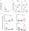
Figure 1
Figure 1
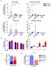
Identification of differentially expressed HH pathway-associated genes in CD34+ CML cells. (A) Volcano plot displaying differential gene expression of 42 HH pathway-associated genes between CD34+ CML patient samples (n = 6) and normal bone marrow (NBM) controls (n = 3) by Bioconductor DESeq2 analysis. Highly differentiated genes are highlighted in red. The size of the dots reflects the mean expression level in absolute read counts and is normalized for sequencing depth. The broken red line indicates the significance threshold (Benjamini-Hochberg-adjusted p < 0.05). (B) Expression levels of the main HH pathway genes between NBM and CML, measured in RPKM. (C) qRT-PCR validation on CD34+ cells from 8 NBM, 8 IM-responders, and 9 IM-nonresponders. Black box: NBM, blue box: IM-responders (IM-R), and rad box: IM-nonresponders (IM-NR). * = p < 0.05; ** = p < 0.01; **** = p < 0.0001.
Figure 2
Figure 2
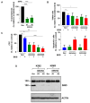
Transcript levels of GLI2 and SMO are increased in LSCs in IM-nonresponders, correlating with their response to SMO inhibition. (A) Relative expression of GLI2 and SMO measured by qRT-PCR in the Lin-CD34+CD38- stem-enriched subpopulation (circles), Lin-CD34+CD38+ progenitor subpopulation (squares), and Lin+CD34- mature hematopoietic subpopulations (triangles) across all CML patient samples (n = 9, left). Expression levels in IM-nonresponders (n = 5, red symbols) and IM-responders (n = 4, blue symbols, right). Each dot represents a single patient sample. (B) Viability assay of CD34+ cells after 72 h of exposure to increasing doses of GL, using the trypan blue exclusion method (n = 3). Apoptosis assay in CD34+ cells after 72 h of treatment with increasing GL doses, using PI and Annexin V staining followed by FACS analysis (n = 3). (C) CFC assay performed on CD34+ CML cells, plated in methylcellulose with growth factors and inhibitors, with colonies counted and typed after 14 days (n = 3). (D) CFC re-plating assay using a portion of colonies from the initial CFC, re-plated into fresh methylcellulose with growth factors but no drugs, with colonies counted and typed after 7–10 days (n = 3). Colony types are indicated by a pattern based on morphology. BFU-E = burst-forming unit-erythroid; CFU-GM = colony-forming unit-granulocyte/macrophage. * = p < 0.05; ** = p < 0.01.
Figure 3
Figure 3
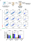
Dual inhibition of SMO and BCR-ABL1 suppresses the growth of CD34+ IM-nonresponder cells in vitro. (A) Viability assay after 72 h using the trypan blue exclusion method. Cells were exposed to GL ± BOS (n = 3). (B) Apoptosis assay after 72 h using Annexin V/PI staining followed by FACS analysis at the same doses used for the viability experiment (n = 3). (C) CFC assay using GL or BOS or a combination (n = 3), followed by a re-plating experiment. (D) LTC-IC-derived CFC measurements were performed on CD34+ CML cells by exposing cells to GL or BOS or both for one week, then conducting half medium changes without drugs for 6 weeks. The cells were subsequently harvested, and a portion was plated into CFC, with colonies counted after 2 weeks. Bar patterns indicate colony types based on morphology. BFU-E = burst-forming unit-erythroid, CFU-GM = colony-forming unit-granulocyte/macrophage. * = p < 0.05; ** = p < 0.01; *** = p < 0.001.
Figure 4
Figure 4
Dual inhibition of SMO and BCR-ABL1 reduces GLI2 expression specifically in LSCs (A,B). This is demonstrated by qRT-PCR analysis of GLI2 and SMO expression in CD34+ subpopulations from three IM-nonresponder patient samples treated with GL or BOS, alone or combined, for 16 h in serum-free medium with or without inhibitors.
Figure 5
Figure 5
Knockdown of SMO in CD34+ stem/progenitor cells reduces cell viability and CFC output. (A) SMO transcript levels in lentiviral-specific SMO shRNA-transduced CD34+ IM-nonresponder cells compared to scramble-transduced cells, measured by qRT-PCR analysis. (B) Viability and apoptosis assays in the same SMO knockdown cells, with or without BOS treatment. (C) CFC assay in these SMO knockdown cells, with or without BOS. (D) SMO protein expression levels in lentiviral-specific SMO shRNA-transduced K562 and K562 IM-resistant (KIMR) cells compared to scramble-transduced cells, measured by Western blot analysis. * = p < 0.05; ** = p < 0.01; *** = p < 0.001.
Figure 6
Figure 6
Combination treatment of BOS and GL reduces engraftment of human CD45+ leukemic cells in mice. (A) Schematic of experimental design using a PDX model. 2.5 × 10⁶ CD34+ IM-nonresponder cells were intravenously injected into each lethally cesium-irradiated NRG mouse, treated with inhibitors, and their engrafted leukemic cells were analyzed. (B) FACS profiles showed engrafted human CD45+ cells in mice BM from each treatment group after one week of oral gavage treatment (3 mice per group). (C) The percentage of human CD45+ cells in BM aspiration from mice of each treatment group was determined at week 10, week 16, and week 29 post-transplantation by FACS analysis. * = p < 0.05; ** = p < 0.01.
Figure 7
Figure 7
Combination treatment of BOS and GL eliminates long-term LSCs in vivo. (A) FACS analysis showing engrafted CD33+CD15+ and CD19+ cells in each mouse across different treatment groups at 10 weeks post-transplantation (left panel). The percentage of human CD33+CD15+ myeloid cells and CD19+ B cells in BM aspiration is shown in the right panel. (B) The percentage of human CD34+ cells in BM from mice in each treatment group at weeks 10, 16, and 29 after transplantation. (C) The percentage of Lin-CD34+CD38- cells in BM aspiration at week 10 post-transplantation. ** = p < 0.01.

References

    1. Rowley J.D. Letter: A new consistent chromosomal abnormality in chronic myelogenous leukaemia identified by quinacrine fluorescence and Giemsa staining. Nature. 1973;243:290–293. doi: 10.1038/243290a0. - DOI - PubMed
    1. Sawyers C.L. Chronic myeloid leukemia. N. Engl. J. Med. 1999;340:1330–1340. doi: 10.1056/NEJM199904293401706. - DOI - PubMed
    1. Baccarani M., Deininger M.W., Rosti G., Hochhaus A., Soverini S., Apperley J.F., Cervantes F., Clark R.E., Cortes J.E., Guilhot F., et al. European LeukemiaNet recommendations for the management of chronic myeloid leukemia: 2013. Blood. 2013;122:872–884. doi: 10.1182/blood-2013-05-501569. - DOI - PMC - PubMed
    1. Druker B.J., Tamura S., Buchdunger E., Ohno S., Segal G.M., Fanning S., Zimmermann J., Lydon N.B. Effects of a selective inhibitor of the Abl tyrosine kinase on the growth of Bcr-Abl positive cells. Nat. Med. 1996;2:561–566. doi: 10.1038/nm0596-561. - DOI - PubMed
    1. Druker B.J., Guilhot F., O'Brien S.G., Gathmann I., Kantarjian H., Gattermann N., Deininger M.W., Silver R.T., Goldman J.M., Stone R.M., et al. Five-year follow-up of patients receiving imatinib for chronic myeloid leukemia. N. Engl. J. Med. 2006;355:2408–2417. doi: 10.1056/NEJMoa062867. - DOI - PubMed

MeSH terms

Grants and funding

LinkOut - more resources