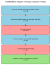
The Role of OCTA and Microperimetry in Revealing Retinal and Choroidal Perfusion and Functional Changes Following Silicone Oil Tamponade in Rhegmatogenous Retinal Detachment: A Narrative Review
- PMID: 41095642
- PMCID: PMC12523867
- DOI: 10.3390/diagnostics15192422
The Role of OCTA and Microperimetry in Revealing Retinal and Choroidal Perfusion and Functional Changes Following Silicone Oil Tamponade in Rhegmatogenous Retinal Detachment: A Narrative Review
Abstract
Background: Rhegmatogenous retinal detachment (RRD), the most common type of retinal detachment, requires prompt surgery to reattach the retina and avoid permanent vision loss. While surgical treatment is adapted to each individual case, one frequent option is pars plana vitrectomy (PPV) with silicone oil (SO) tamponade. Despite achieving anatomical success (complete retinal attachment), concerns persist regarding potential microvascular alterations in the retina and choroid, with a negative impact on visual function. Optical coherence tomography angiography (OCTA) allows detailed, in-depth imaging of retinal and choroidal circulation, whereas microperimetry makes it possible to accurately assess macular function. This review aims to strengthen the existing evidence on vascular and functional alterations at the macular level after SO tamponade in cases of RRD. Methods: A narrative review was conducted using a structured approach, utilizing a PubMed search from January 2000 up to April 2025. Twenty-three studies on OCTA and microperimetry after SO tamponade for RRD were included. Data on vessel densities, choroidal vascular index (CVI), foveal avascular zone (FAZ) size, and retinal sensitivity were extracted and qualitatively analyzed. Results: Studies consistently reported a reduction in the vessel density within the superficial capillary plexus (SCP) under SO tamponade, with partial but incomplete reperfusion post-removal. Choroidal perfusion and CVI were also decreased, exhibiting a negative correlation with the duration of SO tamponade. Microperimetry demonstrated significant reductions in retinal sensitivity (~5-10 dB) during SO tamponade, which modestly improved (~1-2 dB) following removal but generally remaining below normal levels. Conclusions: SO tamponade causes substantial retinal and choroidal vascular impairment and measurable macular dysfunction, even after anatomical reattachment of the retina. It is recommended to perform early SO removal (~3-4 months) and implement routine monitoring by OCTA and microperimetry with the aim of optimizing patient outcomes. Future research should focus on investigating protective strategies and enhancing visual rehabilitation following RRD repair.
Keywords: retinal perfusion; retinal sensitivity; rhegmatogenous retinal detachment; silicone oil tamponade.
Conflict of interest statement
The authors declare that they have no conflicts of interest.
Figures
References
-
- Christou E.E., Stavrakas P., Batsos G., Christodoulou E., Stefaniotou M. Association of OCT-A characteristics with postoperative visual acuity after rhegmatogenous retinal detachment surgery: A review of the literature. Int. Ophthalmol. 2021;41:2283–2292. doi: 10.1007/s10792-021-01777-2. - DOI - PubMed
-
- Mitry D., Charteris D.G., Yorston D., Siddiqui M.A., Campbell H., Murphy A.L., Fleck B.W., Wright A.F., Singh J., Scottish RD Study Group The epidemiology and socioeconomic associations of retinal detachment in Scotland: A two-year prospective population-based study. Investig. Ophthalmol. Vis. Sci. 2010;51:4963–4968. doi: 10.1167/iovs.10-5400. - DOI - PubMed
Publication types
LinkOut - more resources
Full Text Sources
Miscellaneous


