Review
. 2025 Sep 26;15(19):2463.
doi: 10.3390/diagnostics15192463.

Boerhaave Syndrome-Narrative Review

Affiliations
Review

Boerhaave Syndrome-Narrative Review

Dragos Predescu et al. Diagnostics (Basel). .

Abstract

Boerhaave syndrome (BS) and subsequent septic mediastinitis represent a complex cascade of events from esophageal perforation to septic shock. The pathophysiology involves chemical injury, polymicrobial contamination, cytokine storm, endothelial dysfunction, coagulation disorders, and ultimately multiple organ failure. Understanding these mechanisms is crucial for proper therapeutic management that can interrupt this lethal sequence. Due to the complexity of this condition, it is almost impossible to develop a feasible treatment protocol for every situation. The article, through a literature review, evaluates the pathophysiological mechanisms, the consequences of spontaneous esophageal rupture, as well as the therapeutic techniques available for these situations. These elements are the basis of the management of spontaneous esophageal rupture, which involves adapting and customizing the treatment for each patient.

Keywords: Boerhaave syndrome; endoscopic treatment; esophageal perforation; esophageal rupture; mediastinitis.


Conflict of interest statement

The authors declare no conflicts of interest.

Figures

Figure 1
Figure 1
“An Account of the Life and Writings of Herman Boerhaave”—monograph. The smaller image shows Hermann Boerhaave, Doctor of Philosophy and Medicine, Professor of the Theory and Practice of Physic, and of Botany and Chemistry in the University of Leyden; President of the Surgical College in Leyden, Fellow of the Royal Society in London, and Fellow of the Royal Academy in Paris.
Figure 2
Figure 2
The PRISMA diagram.
Figure 3
Figure 3
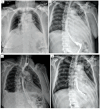
Chest X-ray. Mediastinal aerial image (arrows) is observed that “capes” the esophageal contour, suggestive of an esophageal perforation.
Figure 4
Figure 4
Contrast Swallow Study (water-soluble contrast esophagram). The contrast agent leakage is noted through (A) an esophageal rupture of about 1 cm, and (B) a large esophageal rupture, about 2–3 cm wide by about 7–8 cm long at the level of the middle third.
Figure 5
Figure 5
CT scan of the chest (A) and upper abdomen (B) with contrast agent and cross-sections. Images of a significant hydro-air collection at the mediastinal level (arrows), with extension into the upper abdomen through the esophageal hiatus through an esophageal rupture. Ao—aorta.
Figure 6
Figure 6
Thoraco-abdominal CT scan with contrast agent, sagittal (A) and frontal (B) sections. Images of significant mediastinal, supradiaphragmatic fluid collection (arrows) through an esophageal tear in the middle and lower thirds.
Figure 7
Figure 7
Upper endoscopy images. Different types of lesions due to Boerhaave-type esophageal rupture, with varying sizes and parietal involvement. (A) Esophageal rupture of about 5–6 mm, in the distal esophagus, approximately 5 days after development. (B) Extended esophageal lesion, approximately 2–3 cm wide by approximately 7–8 cm long at the level of the middle third, after 24 h of development. (C) Rupture in the lower esophagus, with necrosis and remnants of esophageal wall, 4 days after the etiological event. (D) Small rupture, 3–4 mm, after an episode of severe vomiting, on the pathological esophagus (eosinophilic esophagitis).
Figure 8
Figure 8
Intraoperative image—open surgery. A large Boerhaave-type esophageal perforation (arrows) is observed, with necrotic margins, with significant mediastinal reaction (remnants of esophageal wall, false membranes), with severe sepsis. The smaller image shows the subtotal esophagectomy specimen.
Figure 9
Figure 9
Post-therapeutic control tomography—cross-section (A). Thoraco-laparoscopic and interventional gastro-enterological surgical multidisciplinary approach, with the evidence of a large hydro-air collection at the mediastinal level and in the right pleural cavity. Note (1) the drain tube (arrow 1) from the pleural collection, (2) the mediastinal drain tube (arrow 2) exteriorized transhiatally and abdominally, and (3) the esophagus with a fully covered stent (arrow 3) with a nasogastric suction probe passed. The smaller image (B) shows the post-procedural stent.
Figure 10
Figure 10
Boerhaave-type esophageal perforation; intraoperative images of minimally invasive surgery in a hybrid treatment. (A) First stage—thoracoscopic approach to hematic pleural collection through mediastinal pleural effusion, with pus, debris and tissue necrosis. (B) Second stage—transhiatal laparoscopic approach to the mediastinal collection.
Figure 11
Figure 11
Post-therapeutic periodic evaluation radiology. (A) Standard radiology 24 h after multidisciplinary intervention, thoraco-laparoscopic surgery and interventional gastroenterology. The stent is noted in the correct position, pleural and mediastinal drainage. (B) Contrast radiology at 5 days, stent in place, no images of esophageal leakage in the mediastinum. (C) Contrast radiology at 14 days, stent well placed, in place, no drainage. (D) Contrast radiology after stent extraction, with normal progression and passage of contrast material, no esophageal leakage.

References

    1. Chirica M., Champault A., Dray X., Sulpice L., Munoz-Bongrand N., Sarfati E., Cattan P. Esophageal perforations. J. Visc. Surg. 2010;147:e117–e128. doi: 10.1016/j.jviscsurg.2010.08.003. - DOI - PubMed
    1. Cuccì M., Caputo F., Fraternali Orcioni G., Roncallo A., Ventura F. Transition of a Mallory-Weiss syndrome to a Boerhaave syndrome confirmed by anamnestic, necroscopic, and autopsy data: A case report. Medicine. 2018;97:e13191. doi: 10.1097/MD.0000000000013191. - DOI - PMC - PubMed
    1. Derbes V.J., Mitchell R.E., Jr. Hermann Boerhaave’s Atrocis, nec descripti prius, morbi historia, the first translation of the classic case report of rupture of the esophagus, with annotations. Bull. Med. Libr. Assoc. 1955;43:217–240. - PMC - PubMed
    1. Haba Y., Yano S., Akizuki H., Hashimoto T., Naito T., Hashiguchi N. Boerhaave syndrome due to excessive alcohol consumption: Two case reports. Int. J. Emerg. Med. 2020;13:56. doi: 10.1186/s12245-020-00318-5. - DOI - PMC - PubMed
    1. Tellechea J.I., Gonzalez J.M., Miranda-García P., Culetto A., D’Journo X.B., Thomas P.A., Barthet M. Role of Endoscopy in the Management of Boerhaave Syndrome. Clin. Endosc. 2018;51:186–191. doi: 10.5946/ce.2017.043. - DOI - PMC - PubMed

LinkOut - more resources