Review
. 2025 Sep 24;17(19):3104.
doi: 10.3390/cancers17193104.

Understanding and Exacerbating the Biological Response of Uveal Melanoma to Proton Beam Therapy

Affiliations
Review

Understanding and Exacerbating the Biological Response of Uveal Melanoma to Proton Beam Therapy

Laura Hawkins et al. Cancers (Basel). .

Abstract

Uveal melanoma (UM) is the most common primary intraocular malignancy in adults, associated with a high tendency for metastasis to the liver. Proton beam therapy (PBT) is the preferred external radiotherapy treatment for primary UM of certain sizes and locations in the eye, due to its efficacy and good local tumour control, as well as its precision to spare surrounding ocular structures. PBT is an effective alternative to surgical enucleation and other non-precision-targeted radiotherapies. Despite this, the radiobiology of UM in response to PBT is still not fully understood. This enhanced knowledge would help to further optimise UM treatment and improve patient outcomes through reducing radiation dosage to ocular structures, treating larger tumours that would otherwise require enucleation, or even offering a treatment strategy for the otherwise fatal liver metastases. In this review, we explore current knowledge of the treatment of UM with PBT, evaluating the biological responses to the therapy. Molecular factors, such as tumour size, oxygen tension levels, DNA damage proficiency, and autophagy, are known to influence the cellular response to radiotherapy, and these will be discussed. Furthermore, we examine innovative strategies to enhance radiotherapy outcomes, such as combination therapies with DNA damage repair and autophagy modulators, as well as advancements in PBT planning and delivery. By integrating current research and emerging technologies, we aim to provide opportunities to improve the therapeutic effectiveness of PBT in UM management.

Keywords: DNA damage; DNA repair; autophagy; hypoxia; ionising radiation; proton beam therapy; uveal melanoma.


Conflict of interest statement

The authors declare no conflicts of interest.

Figures

Figure 1
Figure 1
Depth–dose distribution of PBT relative to the Bragg peak and the relationship to ionisation density/LET. (A) PBT shows a relatively low entrance and exit dose (blue solid line), with the maximal energy being deposited in a well-defined region called the Bragg peak, that can be targeted to the tumour. This is in comparison to X-rays/photons where the dose is highest close to the source (orange line). LET (dashed blue line) increases at and around the Bragg peak. Several proton beams can be modulated and combined to create a spread-out Bragg peak (green line) to target larger tumours. (B) Low-LET radiation tracks cause regions of isolated DNA damage, such as NDA breaks within the tumour cell, whereas more densely ionising high-LET tracks cause regions of CDD. Created in BioRender. Hawkins, L. (2025) https://app.biorender.com/illustrations/6724f4364b0df48f0107bd27?slideId=4ed920ab-b703-4e06-a8cf-d1f8ea75740d (accessed on 13 September 2025).
Figure 2
Figure 2
The major cellular DDR pathways responsive to PBT-induced DNA damage. Ionising radiation induces a variety of DNA lesions that are repaired by DDR pathways. SSBs and base damage are repaired by base excision repair (BER), where damage-specific DNA glycosylases create abasic sites, which are cleaved by APE1, generating SSBs recognised by PARP-1. DNA polymerase β and the XRCC1-DNA ligase IIIα complex fill and seal the gap. DSB repair depends on the cell cycle stage: NHEJ (classical and alternative) is active throughout the cell cycle, with cNHEJ initiated by Ku70/80 binding and recruitment of DNA-PKcs and XRCC4-DNA ligase IV; A-NHEJ involves MRN complex resection, PARP-1 binding, and ligation by DNA ligase I or XRCC1-DNA ligase IIIα. In the S/G2 cell cycle phase, HR repairs DSBs via MRN complex resection, RPA and RAD51 binding, strand invasion by RAD52 and RAD54, and Holliday junction resolution. CDD requires multiple pathways to resolve the lesions, which will depend on the specific nature of the damage. Efficient coordination of these pathways is crucial for the timely repair of PBT-induced damage, promoting cell survival.
Figure 3
Figure 3
Autophagy can have a major influence on cell fate after ionising radiation. In response to radiation, such as PBT, autophagy can be activated to promote either tumour cell survival or death. Protective autophagy can lead to inhibition of apoptosis and the mitigation of oxidative stress, whilst maintaining the integrity of energy homeostasis. Conversely, extensive autophagy, stimulated through either excessive damage or individual cellular preferences, can lead to uncontrolled degradation of essential cellular components, disrupt homeostasis, and trigger cell death either directly through crosstalk with apoptosis, or indirectly through amplifying necroptosis and autophagy-dependent cell death pathways. The dark blue of the cell represents the nucleus, whereas the lighter blue represents the cytoplasm. Image created in BioRender. Hawkins, L. (2025) https://app.biorender.com/illustrations/67373569041045f9a52893c7?slideId=9ffb5c00-6e63-43ff-8466-b9f117882195 (accessed on 13 September 2025).
Figure 4
Figure 4
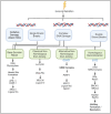
Cellular proteins within the DDR currently under investigation for drug/inhibitors to enhance radiotherapy efficacy. The major repair proteins within the key pathways responsible for co-ordinating the signalling and/or repair of DNA damage induced by PBT are shown. The proteins highlighted in pink have been studied as radiosensitisation targets, and inhibitors have been developed against them with the aim to further increasing the cellular response to radiation. Created in BioRender. Hawkins, L. (2025) https://app.biorender.com/illustrations/662e6ad9a3cc858692ca7beb?slideId=c85d73e2-92cc-40d6-a481-b6f1809d2103 (accessed on 13 September 2025).

References

    1. Tsotridou E., Loukovitis E., Tsiropoulos G.N., Zapsalis K., Pentara I., Tzima K., Eminidou V., Anogeianakis G. Radiation treatment methods in uveal melanoma. Med. Hypothesis Discov. Innov. Ophthalmol. 2021;10:32. doi: 10.51329/mehdiophthal1419. - DOI - PMC - PubMed
    1. Jager M.J., Shields C.L., Cebulla C.M., Abdel-Rahman M.H., Grossniklaus H.E., Stern M.H., Carvajal R.D., Belfort R.N., Jia R., Shields J.A., et al. Uveal melanoma. Nat. Rev. Dis. Primers. 2020;6:24. doi: 10.1038/s41572-020-0158-0. - DOI - PubMed
    1. Khoja L., Atenafu E.G., Suciu S., Leyvraz S., Sato T., Marshall E., Keilholz U., Zimmer L., Patel S.P., Piperno-Neumann S., et al. Meta-analysis in metastatic uveal melanoma to determine progression free and overall survival benchmarks: An international rare cancers initiative (IRCI) ocular melanoma study. Ann. Oncol. 2019;30:1370–1380. doi: 10.1093/annonc/mdz176. - DOI - PubMed
    1. Lane A.M., Kim I.K., Gragoudas E.S. Survival Rates in Patients After Treatment for Metastasis from Uveal Melanoma. JAMA Ophthalmol. 2018;136:981. doi: 10.1001/jamaophthalmol.2018.2466. - DOI - PMC - PubMed
    1. Olofsson Bagge R., Nelson A., Shafazand A., Cahlin C., Carneiro A., Helgadottir H., Levin M., Rizell M., Ullenhag G., Wirén S., et al. A phase Ib randomized multicenter trial of isolated hepatic perfusion in combination with ipilimumab and nivolumab for uveal melanoma metastases (SCANDIUM II trial) ESMO Open. 2024;9:103623. doi: 10.1016/j.esmoop.2024.103623. - DOI - PMC - PubMed

Grants and funding

LinkOut - more resources