. 2025 Oct;29(20):e70852.
doi: 10.1111/jcmm.70852.

Ginsenoside Rg5 Improves Radiation-Induced Heart Injury via PPARG/PDK1/AKT1 Pathway

Affiliations

Ginsenoside Rg5 Improves Radiation-Induced Heart Injury via PPARG/PDK1/AKT1 Pathway

Dao-Ming Zhang et al. J Cell Mol Med. 2025 Oct.

Abstract

Ginsenoside Rg5 (G-Rg5), a rare extract of ginseng, has proven to be valuable for a wide range of clinical applications. However, the role of G-Rg5 in radiation-induced heart injury is currently unclear. This study aims to explore the intervention effect and possible treatment mechanism of G-Rg5 on radiation-induced heart injury. We investigated the impact of G-Rg5 on radiation-induced heart injury through in vivo and in vitro studies. Network pharmacology was employed to explore potential key targets and molecular mechanisms. Various experimental methods were utilised to validate the effects of G-Rg5 on the PPARG/PDK1/AKT1 pathway. G-Rg5 alleviated radiation-induced cardiomyocyte apoptosis and cardiac functional impairment. The expression of peroxisome proliferator activated receptor gamma (PPARG) was upregulated following G-Rg5 treatment, thereby suppressing the transcription of phosphoinositide-dependent protein kinase 1 (PDK1), a predicted target gene regulated by PPARG, to enhance AKT serine/threonine kinase 1 (AKT1) phosphorylation levels. The protective effect of G-Rg5 against radiation-induced heart injury was found to be compromised upon inhibition or knockdown of PPARG. The interaction between PPARG and PDK1 was confirmed by the results of chromatin immunoprecipitation and luciferase reporter assays. G-Rg5, through the upregulation of PPARG expression, induces the transcription of PDK1 and subsequently enhances the phosphorylation levels of AKT1. Ultimately, this process plays a crucial role in mitigating cellular apoptosis and functional decline in radiation-induced heart injury. Therefore, G-Rg5 holds great potential as a therapeutic agent for radiation-induced heart injury.

Keywords: PPARG/PDK1/AKT1; apoptosis; ginsenoside Rg5; radiation‐induced heart disease.


Conflict of interest statement

The authors declare no conflicts of interest.

Figures
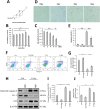
FIGURE 1
FIGURE 1
Ginsenoside Rg5 inhibited radiation‐induced apoptosis of H9c2. (A) Chemical structure of G‐Rg5. (B) Cell viability after 48 h of incubation with different concentrations of G‐Rg5‐containing culture medium. (C) Cell viability after 48 h of radiation in different doses. (D) Representative pictures of cells after different doses of radiation. (E) Bar charts of cell apoptosis rate. (F) Flow cytometry analysis of apoptosis rates in the Con group, Con‐Rg5 group, X‐ray group and X‐Rg5 group. (G–J) Western blot analysis of Cleaved Caspase‐3, BCL‐2 and BAX protein expressions. Data were normalised to the protein expression levels of β‐actin. Data are presented as the mean ± SD per experimental group (n = 6). *p < 0.05, **p < 0.01.
FIGURE 2
FIGURE 2
Ginsenoside Rg5 improved apoptosis and cardiac dysfunctions of RIHD mice. (A) Terminal deoxynucleotidyl transferase‐mediated dUTP nick end labeling (TUNEL) staining (×400 magnification) of mouse myocardial sections. (B) Quantification of TUNEL‐positive cells in heart tissue. (C) Representative M‐mode echocardiographic images of mice. (D) FS, (E) EF, (F, G) ±dP/dt, (H) CO and LVEDP in each group of mice. Data are presented as the mean ± SD per experimental group (n = 3 mice per group). *p < 0.05, **p < 0.01.
FIGURE 3
FIGURE 3
Network pharmacology analysis of Rg5‐RIHD. (A) Venn diagram of shared genes between Rg5 and RIHD. (B) Visual diagram of Ginsenoside Rg5 network ‘Drug Compound‐Targets‐Disease’. (C) Rg5‐RIHD protein interaction network. (D) Top 20 key targets predicted. (E) GO analysis of Rg5‐RIHD shared targets. (F) The Venn diagram prediction of PPARG target genes.
FIGURE 4
FIGURE 4
Binding of ginsenoside Rg5 induced PPARG expression, leading to increased phosphorylation of AKT1. (A) Results of molecular docking shows the interaction between G‐Rg5 and PPARG. Blue sticks represent small molecules, cyan cartoons represent proteins, yellow lines indicate hydrogen bonds and grey dashed lines represent hydrophobic interactions. (B, C) The protein expression of PPARG of CETSA. (D) Representative images of PPARG, PDK1 and p‐AKT1 immunofluorescence in heart tissue. Quantitative results of (E) Bax, (F) Bcl‐2, (G) C‐caspase‐3, (H) p‐AKT1 and (I) PDK1 activity through ELISA detection. Data are presented as the mean ± SD per experimental group. Three independent experiments were conducted for the quantification (E–I). *p < 0.05, **p < 0.01. Scale bar, 100 μm (D).
FIGURE 5
FIGURE 5
Upregulation of PPARG leaded to enhanced PDK1 transcript levels. (A) PPARG binding sites in PDK1 promoter predicted by JASPAR. (B) Schematic images of the potential PPARG binding sites in the promoter of PDK1. (C) ChIP analysis of PPARG occupancy at the PDK1 promoter. (D) Luciferase reporter assays. *p < 0.05, **p < 0.01.
FIGURE 6
FIGURE 6
PPARG knockdown impaired the protective effect of ginsenoside Rg5 on radiation‐induced heart injury. (A) The expression of PPARG mRNA. (B) Western blot analysis of PPARG protein expressions. (C) Bar charts of cell apoptosis rate in the X group, X + Rg5 group, X + siPPARG group and X + Rg5 + siPPARG group. (D) Flow cytometry analysis of apoptosis rates. Quantitative results of (E) C‐caspase‐3, (F) PDK1, (G) p‐AKT1 activity through ELISA detection. Data are presented as the mean ± SD per experimental group (n = 6). *p < 0.05, **p < 0.01.
FIGURE 7
FIGURE 7
Ginsenoside Rg5 improves radiation‐induced heart injury via PPARG/PDK1/AKT1 Pathway.

References

    1. Ellahham S., Khalouf A., Elkhazendar M., Dababo N., and Manla Y., “An Overview of Radiation‐Induced Heart Disease,” Radiation Oncology Journal 40, no. 2 (2022): 89–102. - PMC - PubMed
    1. Ping Z., Peng Y., Lang H., et al., “Oxidative Stress in Radiation‐Induced Cardiotoxicity,” Oxidative Medicine and Cellular Longevity 2020 (2020): 3579143. - PMC - PubMed
    1. Tapio S., “Pathology and Biology of Radiation‐Induced Cardiac Disease,” Journal of Radiation Research 57, no. 5 (2016): 439–448. - PMC - PubMed
    1. Slezak J., Kura B., Babal P., et al., “Potential Markers and Metabolic Processes Involved in the Mechanism of Radiation‐Induced Heart Injury,” Canadian Journal of Physiology and Pharmacology 95, no. 10 (2017): 1190–1203. - PubMed
    1. Kim D. H., “Chemical Diversity of <styled-content style="fixed-case"> Panax ginseng </styled-content>, Panax Quinquifolium, and Panax Notoginseng,” Journal of Ginseng Research 36, no. 1 (2012): 1–15. - PMC - PubMed

MeSH terms

Substances

LinkOut - more resources