Artificial intelligence for diagnosis and prognosis of thymic epithelial tumors: a systematic review
- PMID: 41098391
- PMCID: PMC12518590
- DOI: 10.21037/med-25-34
Artificial intelligence for diagnosis and prognosis of thymic epithelial tumors: a systematic review
Abstract
Background: Thymic epithelial tumors (TETs) are rare, but they are the most common tumors in the anterior mediastinum. The use of artificial intelligence (AI) in the medical field is rapidly advancing. Especially in a rare disease such as TET, AI can help to improve existing care and foster new innovations. However, there are many different AI models with various implications. This systematic review aims to give an overview of recent studies on AI applications for the diagnosis and prognosis of TETs.
Methods: In this systematic review, six electronic databases were searched for research papers with the keywords "thymoma", "thymic epithelial tumors", "thymic carcinoma", "artificial intelligence", "machine learning" and "deep learning". The screening was performed by two reviewers and conflicts were resolved by a third reviewer.
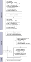
Results: After removal of duplicates, 582 articles were included. Of these, 65 articles were eligible for inclusion after screening. AI models are used mainly for distinguishing between different mediastinal tumor types (n=21), risk stratification (n=26), surgical planning (n=7) and for assisting in subtyping (n=4). For prognostic purposes, AI is used for predicting clinical outcome as well as the chance of metastasis or prognosis (n=7). Models using combined clinical and radiomics data performed better than models with a single type of data. Some of the AI models outperformed physicians or successfully supported physicians in their workflow.
Conclusions: Many AI models have been studied in the context of the diagnosis and prognosis of TETs. Even though results are promising, external validation is often lacking, and sample sizes are small. Therefore, most models are not yet ready for clinical implementation. Further research on AI models with larger datasets and external validation is necessary, with careful consideration of the risk of data leakage.
Keywords: Thymic epithelial tumors (TETs); artificial intelligence (AI); diagnosis; prognosis.
Copyright © 2025 AME Publishing Company. All rights reserved.
Conflict of interest statement
Conflicts of Interest: All authors have completed the ICMJE uniform disclosure form (available at https://med.amegroups.com/article/view/10.21037/med-25-34/coif). “The Series Dedicated to the 14th International Thymic Malignancy Interest Group Annual Meeting (ITMIG 2024)” was commissioned by the editorial office without any funding or sponsorship. J.v.d.T. serves as an unpaid editorial board member of Mediastinum from May 2024 to December 2025. D.D. reported consulting fees from MSD, Amgen, Roche, BMS, Astra Zeneca, and Pfizer. D.D.R. reported grants or contracts from various entities, indicating institutional financial interests without personal financial gain from organizations such as AstraZeneca, BMS, Beigene, Philips, Olink, and Eli Lilly, where his involvement includes research grants, support, and advisory board participation. S.P. reported Hanarth grant financed the INTHYM project, on AI for histopathological classification and recurrence prediction of TET. The authors have no other conflicts of interest to declare.
Figures


References
-
- International Agency for Research on Cancer, World Health Organization, International Academy of Pathology. Thoracic tumours. 5th ed. IARC; 2021.
Publication types
LinkOut - more resources
Full Text Sources
