Editorial
. 2025 Sep 30;14(5):383-388.
doi: 10.21037/acs-2025-evet-26. Epub 2025 Sep 25.

T-NEXT graft: step by step operative technique

Affiliations
Editorial

T-NEXT graft: step by step operative technique

Marco Di Eusanio et al. Ann Cardiothorac Surg. .

Abstract

The frozen elephant trunk (FET) technique has become a cornerstone in the management of complex aortic arch disease, yet reinterventions, both proximally on the root and distally on the thoracoabdominal aorta, remain common. Conventional FET prostheses were designed to recreate standard arch anatomy with the distal anastomosis beyond the left subclavian artery (LSA) and the supra-aortic branches in proximal-to-distal sequence. However, the current trend towards more proximal anastomosis in zones 0-2, brings the arch branches closer to the aortic root, which can limit root access during reoperation by reducing the available clamping zone, and also creates unfavorable angulations for antegrade visceral vessel cannulation during distal endovascular repair. Here, we describe the step-by-step operative technique for a new graft, the T-NEXT, a customized modification of the Thoraflex hybrid prosthesis, designed for improved life-time management of complex aortic disease, featuring a transverse and distal alignment of the arch branches. This configuration leaves an unobstructed proximal graft segment to facilitate safe distal clamping in future proximal reoperations, while preserving a smooth, bidirectional pathway for antegrade and retrograde endovascular access.

Keywords: Frozen elephant trunk (FET); T-NEXT graft; total arch replacement.


Conflict of interest statement

Conflicts of Interest: The authors have no conflicts of interest to declare.

Figures

Figure 1
Figure 1
Extensive dissection of the BCT from its origin to the bifurcation, the left common carotid artery, and the left subclavian artery from its origin to the vertebral artery take-off is carried out. Cardiopulmonary bypass is established by cannulation of the BCT using an interposition 8 mm vascular graft. Venous drainage is achieved through right atrial cannulation, and left ventricular venting is performed via the right superior pulmonary vein. BCT, brachiocephalic trunk.
Figure 2
Figure 2
During the cooling phase, the LSA is ligated proximally, transected, and the proximal stump oversewn. The distal LSA is anastomosed end-to-end to an 8-mm Dacron graft using a running 5-0 polypropylene. This can minimize distal visceral ischemia time and facilitates subsequent LSA reimplantation on the beating heart. This graft is cannulated with a dedicated cannula for antegrade selective cerebral perfusion and connected to the circuit. LSA cannulation is routinely included to enhance cervical spinal cord protection via the vertebral arteries. LSA, left subclavian artery.
Figure 3
Figure 3
During cardioplegia delivery, the left common carotid artery is transected and a dedicated ASCP cannula is inserted to ensure bilateral cerebral perfusion during circulatory arrest. ASCP is delivered at 10 mL/kg/min, equally divided between, equally distributed to the right and left hemispheres, and adjusted to maintain a right radial arterial pressure between 40 and 80 mmHg. Cerebral perfusion is monitored with near-infrared spectroscopy and adjusted accordingly. ASCP, antegrade selective cerebral perfusion.
Figure 4
Figure 4
After reducing the pump flow, the brachiocephalic trunk is clamped, and the aortic cross-clamp removed. The ascending aorta and the proximal aortic arch (up to the target landing zone) are resected.
Figure 5
Figure 5
The stent-graft is gently bent to match the curvature of the descending thoracic aorta and stabilized with the delivery handle. A black orientation marker, aligned with the brachiocephalic trunk branch, ensures accurate positioning. The release sequence is identical to that of a conventional Thoraflex graft: the sheath is retracted through the splitter to deploy the self-expanding stent, the splitter is removed, the sewing collar freed and the stent tip released by removing the central locking pin.
Figure 6
Figure 6
Distal aortic anastomosis. The sewing collar of the T-NEXT graft is sutured to the distal aortic arch stump using a running 3-0 or 4-0 polypropylene suture. A circumferential Teflon felt strip is used for reinforcement.
Figure 7
Figure 7
After completion of the distal anastomosis, a perfusion cannula is inserted into the T-NEXT side branch, the graft is de-aired, and antegrade distal organ perfusion is initiated. Hemostasis is checked, and systemic rewarming is started. The proximal anastomosis to the ascending aorta is performed with a running 4-0 polypropylene suture, and the heart reperfused.
Figure 8
Figure 8
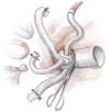
On the beating heart, the interposition left subclavian artery graft is anastomosed end-to-end to one limb of the bifurcated branch of the T-NEXT graft using a running 5-0 polypropylene suture.
Figure 9
Figure 9
The left common carotid artery is reimplanted onto the second limb of the bifurcated branch, while the brachiocephalic trunk is reimplanted end-to-end onto the single dedicated branch of the T-NEXT graft.
Figure 10
Figure 10
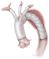
Schematic of the T-NEXT graft configuration. The graft features a transverse distal alignment of branches: a bifurcated branch for the LSA and LCCA, and a single branch for the BCT, each oriented at 90° to the graft body. This design leaves the proximal graft segment unobstructed, facilitating distal clamping in future reoperations and preserving a bidirectional catheter pathway between upper and lower body vessels for subsequent endovascular interventions. BCT, brachiocephalic trunk; LCCA, left common carotid artery; LSA, left subclavian artery.

References

    1. Di Eusanio M, Gatta E. T-next: a new custom-made Thoraflex graft to simplify proximal and distal aortic reinterventions. Eur J Cardiothorac Surg 2023;63:ezad232. 10.1093/ejcts/ezad232 - DOI - PubMed
    1. Ma WG, Zheng J, Sun LZ, et al. Open Stented Grafts for Frozen Elephant Trunk Technique: Technical Aspects and Current Outcomes. Aorta (Stamford) 2015;3:122-35. 10.12945/j.aorta.2015.14.062 - DOI - PMC - PubMed
    1. Berretta P, Galeazzi M, Malvindi PG, et al. Frozen elephant trunk with T-NEXT. Ann Cardiothorac Surg 2025;14:314-5. 10.21037/acs-2025-evet-0017 - DOI - PMC - PubMed
    1. Berretta P, Galeazzi M, Malvindi PG, et al. Frozen elephant use in type a dissection: fundamentals, innovations, and pitfalls. Expert Rev Med Devices 2024;21:579-86. 10.1080/17434440.2024.2365416 - DOI - PubMed

Publication types