

Immunopathological studies of orthotopic human liver allografts
- PMID: 4109928
- PMCID: PMC2982185
- DOI: 10.1016/s0140-6736(72)90288-7
Immunopathological studies of orthotopic human liver allografts
Abstract
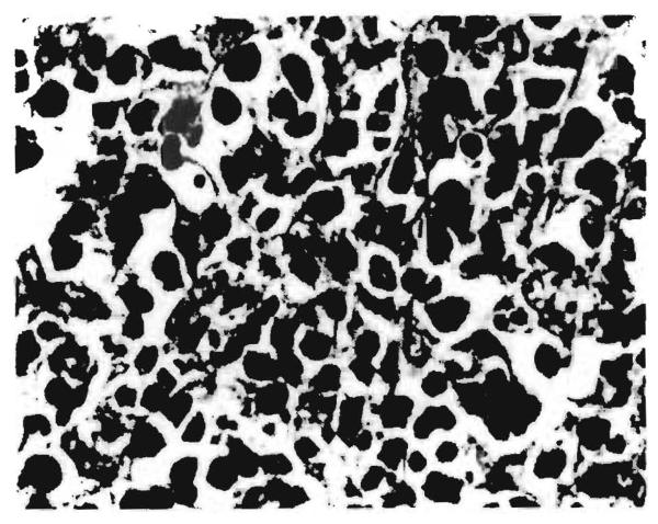
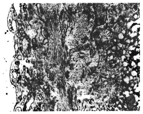
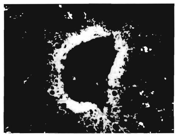
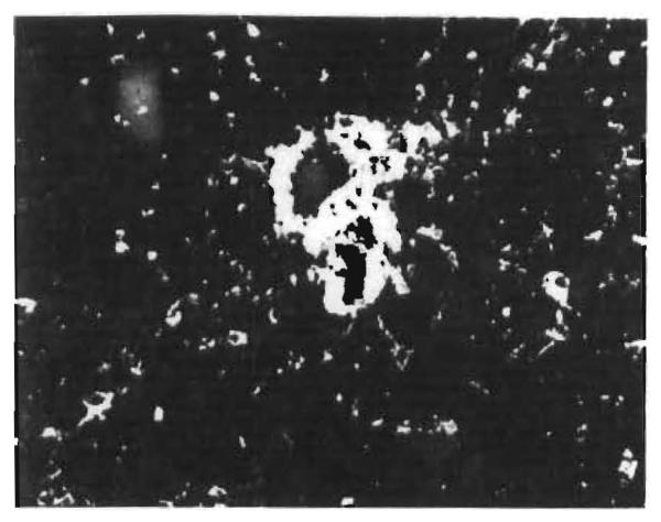
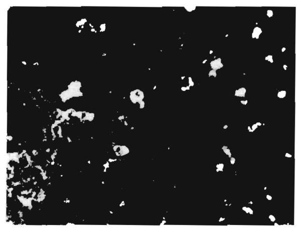
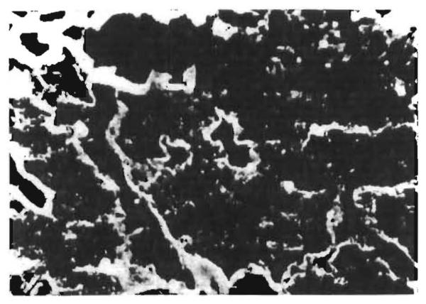
Twenty-six specimens obtained from twenty human orthotopic liver allografts 10–968 days after transplantation were studied by light microscopy, electron microscopy, and immunofluorescence. The main lesions consisted of mononuclear-cell infiltration around the portal tracts, centrilobular cholestasis, liver-cell atrophy and reticulin collapse, obliterative intimal thickening of hepatic arteries, and fibrosis. Moderate amounts of IgG and/or IgM and complement (β1C/β1A globulin or C'lq) were observed in four of the liver samples and smaller deposits were present in another five. A further three specimens contained IgG without complement. IgA was detected in only one of the samples. The immunoglobulins were found in the walls of the portal and central veins and of the sinusoids in all thirteen positive liver samples, in the walls of branches of the hepatic artery in three, and in the cytoplasm of some of the mononuclear cells infiltrating the portal tracts in nine of the specimens. Fibrinogen was seen in eight of the samples, usually in the spaces of Disse. Accumulations of immunoglobulins and complement were less frequent in liver than in kidney and heart allografts. These findings suggest that in the failure of human liver allografts cell-mediated immunity and non-immunological factors may be more important than humoral antibody.
Figures
References
MeSH terms
Substances
Grants and funding
LinkOut - more resources
Full Text Sources
Miscellaneous
