. 2025 Oct 1:16:1678724.
doi: 10.3389/fphar.2025.1678724. eCollection 2025.

Ferulic acid mitigates 3-Nitropropionic acid-induced Huntington's disease via modulation of Nrf2/HO-1, TLR4/NF-κB, and SIRT1/p53 signaling pathways

Affiliations

Ferulic acid mitigates 3-Nitropropionic acid-induced Huntington's disease via modulation of Nrf2/HO-1, TLR4/NF-κB, and SIRT1/p53 signaling pathways

Mohamed A Abdelgawad et al. Front Pharmacol. .

Abstract

Background: Ferulic acid (FA) is a natural phenolic compound that has demonstrated effectiveness against Huntington's disease (HD). However, its exact mechanism remains unclear. Therefore, the current study aims to investigate FA's potential mechanism of action against 3-nitropropionic acid (3NP)-induced HD.

Methods: Adult male Wistar albino rats were administered FA orally (100 mg/kg) for 3 weeks, and 3NP (10 mg/kg) was intraperitoneally administered during the last 2 weeks to induce HD. Behavioral performance was assessed using the open field and hanging wire tests. Striatal tissue was analyzed using ELISA, qRT-PCR, Western blotting, histopathology, and immunohistochemistry.

Results: Administration of 3NP led to weight loss, neurobehavioral deficits, oxidative damage, apoptotic cell death, and neuroinflammation. FA treatment mitigated these pathological changes by activating Nrf2/HO-1 signaling, a critical player in cellular redox balance. This beneficial effect was mirrored in restoring TAC levels and suppressing MDA. Moreover, FA suppressed TLR4/NF-κB inflammatory signaling, thereby reducing TNF-α and IL-1β levels. In addition, the anti-apoptotic properties of FA were confirmed by modulating SIRT1/p53 signaling, leading to Bcl-2 enhancement and caspase-3 downsizing. Furthermore, FA enhanced neuronal survival and plasticity confirmed by neurotrophic BDNF elevation. Histopathological and immunohistochemical analyses confirmed improved neuronal survival and reduced gliosis following FA treatment.

Conclusion: The current research demonstrates that FA exhibits potent neuroprotective effects in experimental HD by modifying Nrf2/HO-1, TLR4/NF-κB, and SIRT1/p53 signaling pathways. These findings provide new mechanistic insights into FA's potential role in managing HD.

Keywords: 3-Nitropropionic acid; Nrf2; SIRT1; TLR4; ferulic acid; neuroinflammation.


Conflict of interest statement

The authors declare that the research was conducted in the absence of any commercial or financial relationships that could be construed as a potential conflict of interest.

Figures

FIGURE 1
FIGURE 1
Schematic presentation of experimental design.
FIGURE 2
FIGURE 2
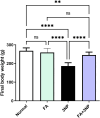
FA intake impacts on the final body weight in rats intoxicated with 3NP. Data are presented as mean ± SD; ns (non-significant, P > 0.05), *P < 0.05, **P < 0.01, ***P < 0.001, and ****P < 0.0001.
FIGURE 3
FIGURE 3
FA intake impacts on motor parameters (a), representative movement tracks; (b), distance traveled; (c), speed average; (d), immobility time and (e) fall of latency in rats intoxicated with 3NP. Data are presented as mean ± SD; ns (non-significant, P > 0.05), *P < 0.05, **P < 0.01, ***P < 0.001, and ****P < 0.0001.
FIGURE 4
FIGURE 4
FA intake impacts on striatal (a) Nrf2 and (b) HO-1 mRNA expression, as well as (c) TAC and (d) MDA content in rats subjected to 3NP. Data are presented as mean ± SD; ns (non-significant, P > 0.05), *P < 0.05, **P < 0.01, ***P < 0.001, and ****P < 0.0001.
FIGURE 5
FIGURE 5
FA intake impacts on striatal neurotrophic BDNF content in rats subjected to 3NP. Data are presented as mean ± SD; ns (non-significant, P > 0.05), *P < 0.05, **P < 0.01, ***P < 0.001, and ****P < 0.0001.
FIGURE 6
FIGURE 6
FA intake impacts on striatal (a) TLR4 mRNA expression, (b) NF-κB p65 IHC protein expression, as well as protein content of (c) TNF-α and (d) IL-1β in rats subjected to 3NP. Data are presented as mean ± SD; ns (non-significant, P > 0.05), *P < 0.05, **P < 0.01, ***P < 0.001, and ****P < 0.0001.
FIGURE 7
FIGURE 7
FA intake impacts on striatal (a) p53, (b) Bcl-2 content, and (c) caspase-3 IHC in rats subjected to 3NP. Data are presented as mean ± SD; ns (non-significant, P > 0.05), *P < 0.05, **P < 0.01, ***P < 0.001, and ****P < 0.0001.
FIGURE 8
FIGURE 8
FA intake impacts on striatal protein expressions of SIRT1 in rats subjected to 3NP. Data are presented as mean ± SD; ns (non-significant, P > 0.05), *P < 0.05, **P < 0.01, ***P < 0.001, and ****P < 0.0001.
FIGURE 9
FIGURE 9
Representative photomicrographs of H & E stained stratia in the normal and FA groups showing intact neurons with normal histological architecture. 3NP group, showing gliosis (black arrow), distinct capillaries (green arrow), and edema (red arrow). FA+3NP group, showing apparently normal striatum.
FIGURE 10
FIGURE 10
FA intake impacts on the neuronal survival rate in rats exposed to 3NP. Normal and FA groups, showing apparently normal lightly stained neurons within the striatum. 3NP group, showing numerous dark degenerating neurons (arrows) within the striatum. FA+3NP group, few dark degenerating neurons (arrows) within the striatum. Data are presented as mean ± SD; ns (non-significant, P > 0.05), *P < 0.05, **P < 0.01, ***P < 0.001, and ****P < 0.0001.
FIGURE 11
FIGURE 11
FA intake impacts on striatal GFAP in rats exposed to 3NP. Photomicrograph of brain, striatum, Normal and FA groups showing normal mild GFAP expression (Immune staining), 3NP group showing intense GFAP expression, and FA+3NP group showing moderate GFAP expression. Data are presented as mean ± SD; ns (non-significant, P > 0.05), *P < 0.05, **P < 0.01, ***P < 0.001, and ****P < 0.0001.

References

    1. Ahmed L. A., Darwish H. A., Abdelsalam R. M., Amin H. A. (2016). Role of rho kinase inhibition in the protective effect of fasudil and simvastatin against 3-nitropropionic acid-induced striatal neurodegeneration and mitochondrial dysfunction in rats. Mol. Neurobiol. 53, 3927–3938. 10.1007/s12035-015-9303-2 - DOI - PubMed
    1. Ayala-Peña S. (2013). Role of oxidative DNA damage in mitochondrial dysfunction and Huntington’s disease pathogenesis. Free Radic. Biol. Med. 62, 102–110. 10.1016/j.freeradbiomed.2013.04.017 - DOI - PMC - PubMed
    1. Azman K. F., Zakaria R. (2025). Brain-derived neurotrophic factor (BDNF) in Huntington’s disease: neurobiology and therapeutic potential. Curr. Neuropharmacol. 23, 384–403. 10.2174/1570159X22666240530105516 - DOI - PMC - PubMed
    1. Bono-Yagüe J., Gómez-Escribano A. P., Millán J. M., Vázquez-Manrique R. P. (2020). Reactive species in Huntington disease: are they really the radicals you want to catch? Antioxidants 9, 577. 10.3390/antiox9070577 - DOI - PMC - PubMed
    1. Burnette W. N. (1981). “Western blotting”: electrophoretic transfer of proteins from sodium dodecyl sulfate-polyacrylamide gels to unmodified nitrocellulose and radiographic detection with antibody and radioiodinated protein A. Anal. Biochem. 112, 195–203. 10.1016/0003-2697(81)90281-5 - DOI - PubMed

LinkOut - more resources