. 2025 Oct 17;25(1):359.
doi: 10.1186/s12935-025-04008-1.

Suppression of pentraxin 3 inhibits human osteosarcoma cell metastasis by repressing MAZ through STAT3 pathway

Affiliations

Suppression of pentraxin 3 inhibits human osteosarcoma cell metastasis by repressing MAZ through STAT3 pathway

Chia-Hsuan Chou et al. Cancer Cell Int. .

Abstract

Background: Pentraxin 3 (PTX3), also known as TNF-inducible gene 14 protein (TSG-14), exerts pleiotropic roles in inflammation, immune responses, and a myriad of cancers, but there is limited knowledge regarding the role of PTX3 in the metastasis of human osteosarcoma.

Methods: Using RNA sequencing technology, transfection, western blotting, flow cytometry, and luciferase reporter, colony formation, quantitative reverse transcription–polymerase chain reaction (qRT-PCR), boyden chamber assays, and a xenograft model, the effects of PTX3 on cell migration and invasion in human osteosarcoma cells were investigated.

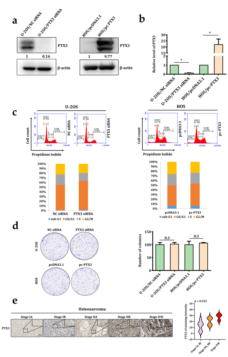
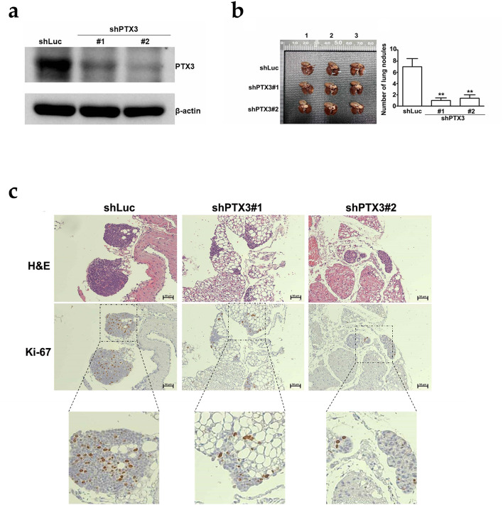
Results: PTX3 silencing repressed migration and invasion in U-2OS cells and PTX3 overexpression accelerated migration and invasion in HOS cells. Recombinant human PTX3 (rhPTX3) increased migration and invasion of HOS cells. In PTX3 knockdown U-2OS cells, the heatmap revealed the downregulation of Myc-associated zinc finger protein (MAZ), which is associated with the epithelial-mesenchymal transition pathway and osteosarcoma tissues. MAZ overexpression rescued the inhibition of migration by PTX3 knockdown in osteosarcoma cells. The activity of the MAZ promoter was reduced with PTX3 knockdown in osteosarcoma cells. While Stat3 overexpression rescued the inhibition of MAZ expression and migration by PTX3 knockdown in osteosarcoma cells. Moreover, in tail vein injection experiments, shPTX3 U-2OS cells reduced pulmonary metastasis formation in immunodeficient nude mice. By repressing MAZ through Stat3, suppression of PTX3 inhibits migration and invasion in osteosarcoma cells, and knockdown of PTX3 reduces pulmonary metastasis.

Conclusions: Our results raise interest in further study of PTX3’s anti-metastasis properties in osteosarcoma, providing molecular insight into PTX3’s role in osteosarcoma metastasis.

Supplementary Information: The online version contains supplementary material available at 10.1186/s12935-025-04008-1.

Keywords: Invasion; MAZ; Migration; Osteosarcoma; Pentraxin 3.


Conflict of interest statement

Declarations. Ethics approval and consent to participate: Not applicable. Consent for publication: Not applicable. Competing interests: The authors declare no competing interests.

Figures

Fig. 1
Fig. 1
PTX3 expression and functional evaluation in osteosarcoma. (a) Total lysates obtained from U-2OS and HOS osteosarcoma cells were transfected with NC siRNA, PTX3 siRNA or pcDNA3.1, pc-PTX3 plasmid, and subsequently detected by western blotting. (b) Relative of PTX3 expression in U-2OS and HOS cell lines transfected with NC siRNA, PTX3 siRNA or pcDNA3.1, pc-PTX3 plasmid were detected by qRT-PCR assay. Flow cytometry analysis (c), colony formation (d) in U-2OS and HOS cells transfected with NC siRNA, PTX3 siRNA or pcDNA3.1, pc-PTX3 plasmid. (e) IHC staining of PTX3 in a human osteosarcoma tissue array. Representative images show PTX3 expression across different osteosarcoma clinical stages (scale bar: 20 μm), then quantified. NC, Negative control; pc-PTX3, pcDNA3.1-PTX3; qRT-PCR, quantitative real-time polymerase chain reaction. The data are presented as mean ± standard deviations. *p < 0.05 compared with control cells
Fig. 2
Fig. 2
Suppression of PTX3 inhibits the migration and invasion of human osteosarcoma cells in vitro. U-2OS (a) and HOS (b) osteosarcoma cells transfected with NC siRNA, PTX3 siRNA or pcDNA3.1, pc-PTX3 plasmid were collected to determine the cell migration and invasion capabilities. U-2OS and HOS osteosarcoma cells were treated with recombinant PTX3 protein, and subsequently collected for the assessment of cell migration (c) and invasion (d). NC, Negative control; pc-PTX3, pcDNA3.1-PTX3. The data are presented as mean ± standard deviations. *p < 0.05 compared with control cells. ANOVA analysis with Tukey’s posteriori comparison was used. a Significantly different, p < 0.05 when compared to control. b Significantly different, p < 0.05, when compared to 50 ng/mL
Fig. 3
Fig. 3
PTX3 regulates downstream MAZ. (a) A volcano plot illustrates the distribution of all differentially expressed gene, a |log2 fold change| ≧ 1 and p value < 0.05 are considered as significant differences in U-2OS cells transfected with PTX3 siRNA and NC siRNA. Mapping the top of 20 upregulated genes and downregulated genes. Red dots represent significantly upregulated genes, while blue dots represent significantly downregulated genes. (b) Visualization of RNA-Seq differential expression data with heatmap. qRT-PCR was used for detecting EMT-related gene in U-2OS (c) and HOS (d) cell lines that transfected with NC siRNA, PTX3 siRNA or pcDNA3.1, pc-PTX3 plasmid. The expression of PTX3, RPIA, GLO1, and MAZ in U-2OS (e) and HOS (f) cell lines that transfected with NC siRNA, PTX3 siRNA or pcDNA3.1, pc-PTX3 plasmid were detected by western blotting. (g) The correlation between of PTX3 and MAZ expression in sarcoma cell lines (n = 68). NC, Negative control; pc-PTX3, pcDNA3.1-PTX3; qRT-PCR, quantitative real-time polymerase chain reaction. The data are presented as mean ± standard deviations. *p < 0.05 compared with control cells
Fig. 4
Fig. 4
Suppression of PTX3 downregulates MAZ to inhibit migration and invasion of osteosarcoma cells. The efficiency of MAZ knockdown/overexpression in U-2OS and HOS cell lines transfected with NC siRNA, MAZ siRNA or pCMV6, pCMV6-MAZ were detected by Western blotting (a) and qRT-PCR (b). U-2OS (c) and HOS (d) osteosarcoma cells transfected with NC siRNA, MAZ siRNA or pCMV6, pCMV6-MAZ plasmid were collected to determine the cell migration and invasion capabilities. U-2OS (e) and HOS (f) osteosarcoma cells transfected with NC siRNA, PTX3 siRNA or pcDNA3.1, pc-PTX3 plasmid in MAZ promoter activity were collected to determine MAZ promoter reporter assay. (g) Following the downregulation of PTX3, we overexpressed MAZ in U-2OS cells to assess MAZ expression and cell migration. *p < 0.05 compared with control cells. #p < 0.05 compared with MAZ-overexpressed group. (h) Following the overexpression of PTX3, we knocked down MAZ in HOS cells to assess MAZ expression and cell migration. NC, negative control; qRT-PCR, quantitative real-time polymerase chain reaction. The data are presented as mean ± standard deviations. *p < 0.05 compared with control cells. #p < 0.05 compared with MAZ siRNA group
Fig. 5
Fig. 5
Suppression of PTX3 downregulates MAZ through repressing STAT3 to inhibit migration of osteosarcoma cells. U-2OS (a) and HOS (b) osteosarcoma cells transfected with NC siRNA, PTX3 siRNA or pcDNA3.1, pc-PTX3 plasmid were collected for the detection of FAK, MAPKs, Akt, and Stat3 pathways expression through western blotting. Following the downregulation of PTX3, we overexpressed Stat3 in U-2OS cells to assess MAZ expression (c) and cell migration (e). *p < 0.05 compared with control cells. #p < 0.05 compared with Stat3-overexpressed group. Following the overexpression of PTX3, we treatment with C188-9 (30 µM) in HOS cells to assess MAZ expression (d) and cell migration (f). NC, negative control; pc-PTX3, pcDNA3.1-PTX3; pc-Stat3, pcDNA3.0-Stat3. *p < 0.05 compared with control cells. #p < 0.05 compared with C188-9-treated group. The data are presented as mean ± standard deviations
Fig. 6
Fig. 6
Downregulation of PTX3 inhibits pulmonary metastasis in vivo. (a) Cells were infected with shLuc and shPTX3 and then analyzed by western blotting. (b) The lungs of mice in both the shLuc group and the shPTX3 group exhibit metastatic nodules. (c) The lungs were fixed in 4% paraformaldehyde, sectioned, and stained with H&E and IHC using anti-Ki-67. H&E, Hematoxylin and Eosin. The data are presented as mean ± standard deviations. *p < 0.05 compared with shLuc cells
Fig. 7
Fig. 7
A schematic diagram illustrates that knockdown of PTX3 inhibits MAZ expression through phosphorylation of the Stat3 pathway and reduces the potential for lung metastasis in osteosarcoma cells

References

    1. Lu KH, Lu EW, Lin CW, Yang JS, Yang SF. New insights into molecular and cellular mechanisms of zoledronate in human osteosarcoma. Pharmacol Ther. 2020;214:107611. - PubMed
    1. Otoukesh B, Abbasi M, Gorgani HO, Farahini H, Moghtadaei M, Boddouhi B, et al. Micrornas signatures, bioinformatics analysis of miRNAs, miRNA mimics and antagonists, and miRNA therapeutics in osteosarcoma. Cancer Cell Int. 2020;20:254. - PMC - PubMed
    1. Rathore R, Van Tine BA. Pathogenesis and current treatment of osteosarcoma: perspectives for future therapies. J Clin Med. 2021. 10.3390/jcm10061182 - PMC - PubMed
    1. Smrke A, Anderson PM, Gulia A, Gennatas S, Huang PH, Jones RL. Future directions in the treatment of osteosarcoma. Cells. 2021. 10.3390/cells10010172 - PMC - PubMed
    1. Fernandes I, Melo-Alvim C, Lopes-Bras R, Esperanca-Martins M, Costa L. Osteosarcoma pathogenesis leads the way to new target treatments. Int J Mol Sci. 2021. 10.3390/ijms22020813 - PMC - PubMed

LinkOut - more resources