. 2025 Oct 17;23(1):1124.
doi: 10.1186/s12967-025-07168-w.

IL-17 + γδT cell: a new target in hyperbaric oxygen treatment reducing spinal cord injury

Affiliations

IL-17 + γδT cell: a new target in hyperbaric oxygen treatment reducing spinal cord injury

Mo Liu et al. J Transl Med. .

Abstract

Background: Hyperbaric oxygen (HBO) treatment can attenuate the inflammatory response after spinal cord injury (SCI), but the specific molecular mechanisms are unclear. γδT cells have emerged as key immune regulatory cells. This study aims to analyze the mechanism of HBO treatment reducing SCI mainly from the perspective of γδT cells.

Methods: Contusion SCI models were established in WT, TCRδ-/-, and IL-17-/- mice, and HBO treatment was performed. Hindlimb locomotor function, ChAT-positive motor neuron numbers, γδT cell proportion, and inflammatory cytokines level of different experimental groups were compared after SCI. Furthermore, we measured the protein and gene expression of STAT3 and RORγt in both WT mice and STAT3 overexpression mice following HBO treatment.

Results: Our results showed γδT cells, especially IL-17 + γδT cells, were involved in SCI. HBO treatment improved locomotor recovery, reduced the proportion of γδT and IL-17 + γδT cells, and decreased inflammatory cytokines in WT mice after SCI. However, HBO treatment did not affect locomotor recovery or inflammatory cytokines level in TCRδ-/- or IL-17-/- mice after SCI. In addition, HBO treatment significantly inhibited the expression of STAT3 and RORγt in WT mice after SCI. Over-expression of STAT3 weakened the inhibitory effect of HBO on IL-17 + γδT cells and inflammatory cytokines after SCI.

Conclusions: These findings indicated that IL-17 + γδT cells may play a key role in HBO treatment alleviating the inflammatory response after SCI. HBO treatment may regulate IL-17 + γδT cells by modulating the pathway of STAT3/RORγt. This study provides the theoretical basis and potential therapeutic target for HBO treatment in SCI.

Keywords: Hyperbaric oxygen; Inflammatory cytokines; Spinal cord injury; γδT cell.


Conflict of interest statement

Declarations. Ethics approval and consent to participate: All experimental procedures were conducted with the approval of the Ethics Committee of Beijing Chao-Yang Hospital, Capital Medical University (approval No. 2021-3-1-26). Consent for publication: All authors gave their consent for publication. Competing interests: The authors declare that there are no competing interests associated with the manuscript.

Figures

Fig. 1
Fig. 1
HBO treatment contributes to locomotor recovery and motor neuron recovery of mice with SCI. A Using MASCIS III, a 10 g rod was released from a distance of 6.25 mm to inflict the exposed dura mater at T10 (left diagram). The right diagram shows an example of spinal cord tissue instantly after inflict; B BMS test to show hindlimb locomotor function recovery from 0 to 49dpi (n ≥ 6); C Representative images of immunofluorescence staining of ChAT (red). Nuclei were labeled with DAPI (blue). Scale bar: 200 μm (left), 50 μm (right). D Quantification of the relative fluorescence intensity of the ChAT (n = 4). Statistics: Data represent mean ± SD, ***P < 0.001, ****P < 0.0001. B Two-way repeated measures ANOVA with Bonferroni correction test; D One-way ANOVA with Tukey's post-hoc test. SH: sham, SCI: spinal cord injury, HBO: hyperbaric oxygen
Fig. 2
Fig. 2
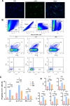
HBO treatment reduces γδT cell proportion and inflammatory cytokines level in mice with SCI. A Representative images of immunofluorescence staining of TCR (Green). Nuclei were labeled with DAPI (blue). Scale bar: 20um. B The gating strategy (Cells → Singlets → Live cells → CD45 + cells). Representative images of flow cytometry of γδT (gate on CD45 + cells), IL-17 + γδT, and IFN-γ + γδT cells (gate on CD3 + γδTCR + cells) in the damaged spinal cord tissues of SH, SCI, and HBO groups. C Quantitative analysis of the proportion of γδT, IL-17 + γδT, and IFN-γ + γδT cells in the damaged spinal cord tissues of three groups (n = 6). D Comparison of the expression levels of IL-17, IFN-γ, IL-1β, IL-6, and TNF-α in three groups (n = 6). Statistics: Data represent mean ± SD, *P < 0.05, **P < 0.01, *** P < 0.001, **** P < 0.0001, ns: no significance. C, D One-way ANOVA with Tukey's post-hoc test. SH: sham, SCI: spinal cord injury, HBO: hyperbaric oxygen
Fig. 3
Fig. 3
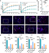
Comparison of BMS scores, ChAT relative fluorescence intensity, and inflammatory cytokines level in TCRδ −/− mice versus WT mice. A BMS test to show hindlimb locomotor functional recovery from 0 to 49 dpi in four groups. B The image analysis results were presented as the relative intensity of the fluorescence of ChAT in four groups (n = 4). C Representative images of immunofluorescence staining of ChAT (red). Nuclei were labeled with DAPI (blue). Scale bar: 100 μm (left), 50 μm (right). D Comparison of the expression levels of IL-17, IFN-γ, IL-1β, IL-6, and TNF-α in four groups (n = 6). Statistics: Data represent mean ± SD, * P < 0.05, ** P < 0.01, *** P < 0.001, **** P < 0.0001, ns: no significance. A Two-way repeated measures ANOVA combined with Bonferroni correction test; B, D One-way ANOVA with Tukey's post-hoc test. SH: sham, SCI: spinal cord injury, HBO: hyperbaric oxygen
Fig. 4
Fig. 4
Comparison of BMS scores, ChAT relative fluorescence intensity, and inflammatory cytokines level in IL-17−/− mice versus WT mice. A BMS test to show hindlimb locomotor functional recovery from 0 to 49dpi in four groups (n ≥ 6); B The image analysis results were presented as the relative fluorescence intensity of ChAT in four groups (n = 4); C Representative images of immunofluorescence staining of ChAT (red). Nuclei were labeled with DAPI (blue). Scale bar: 100 μm (left), 50 μm (right); D Comparison of the expression levels of IFN-γ, IL-1β, IL-6, and TNF-α in four groups (n = 6). Statistics: Data represent mean ± SD, * P < 0.05, *** P < 0.001, **** P < 0.0001, ns: no significance. A Two-way repeated measures ANOVA combined with Bonferroni correction test; B, D One-way ANOVA with Tukey's post-hoc test. SH: sham, SCI: spinal cord injury, HBO: hyperbaric oxygen
Fig. 5
Fig. 5
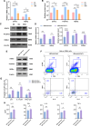
HBO treatment regulates the expression of STAT3 and RORγt in mice with SCI. A mRNA levels of STAT3 and RORγt in SH, SCI, and HBO group (n = 6); B Protein expression levels of STAT3, p-STAT3, and RORγt in three groups (n = 4); C Representative Western blotting results of STAT3, P-STAT3, and RORγt within spinal cords harvested 7dpi in three groups; D mRNA levels of STAT3 and RORγt in HBO + AAV-Null and HBO + AAV-STAT3 group (n = 6), and protein expression levels of STAT3, p-STAT3, and RORγt in two groups (n = 4); E Representative Western blotting results of STAT3, P-STAT3, and RORγt within spinal cords harvested 7dpi in two groups; F The gating strategy (Cells → Singlets → Live cells → CD45 + cells). Representative images of flow cytometry of γδT (gate on CD45 + cells), IL-17 + γδT, and IFN-γ + γδT cells (gate on CD3 + γδTCR + cells) in the damaged spinal cord tissues of two groups. G Quantitative analysis of the proportion of γδT, IL-17 + γδT, and IFN-γ + γδT cells in the damaged spinal cord tissues of two groups (n = 6). H Comparison of the expression levels of IL-17, IFN-γ, IL-1β, IL-6, and TNF-α in two groups (n = 6). Statistics: Data represent mean ± SD, *P < 0.05, ** P < 0.01, *** P < 0.001, **** P < 0.0001, ns: no significance. A, B One-way ANOVA with Tukey's post-hoc test; D, G, H Independent samples t-tests. SH: sham, SCI: spinal cord injury, HBO: hyperbaric oxygen
Fig. 6
Fig. 6
Mechanism of HBO treatment regulating IL-17 + γδT cells to alleviate SCI. HBO treatment may regulate IL-17 + γδT cells by inhibiting STAT3/RORγt pathway. HBO treatment could reduce the proliferation and differentiation of IL-17 + γδT cells and inhibit the secretion of inflammatory cytokines, which reduces the inflammatory response after SCI and promotes the recovery of locomotor function in SCI mice. (The figure was created using figdraw.com.)

References

    1. Carelli S, Giallongo T, Rey F, Colli M, Tosi D, Bulfamante G, et al. Neuroprotection, recovery of function and endogenous neurogenesis in traumatic spinal cord injury following transplantation of activated adipose tissue. Cells. 2019;8(4):329. - PMC - PubMed
    1. Guan B, Anderson DB, Chen L, Feng S, Zhou H. Global, regional and national burden of traumatic brain injury and spinal cord injury, 1990–2019: a systematic analysis for the Global Burden of Disease Study 2019. BMJ Open. 2023;13(10):e075049. - PMC - PubMed
    1. Quadri SA, Farooqui M, Ikram A, Zafar A, Khan M, Suriya SS, et al. Recent update on basic mechanisms of spinal cord injury. Neurosurg Rev. 2020;43(2):425–41. - PubMed
    1. He X, Deng B, Ma M, Wang K, Li Y, Wang Y, et al. Bioinformatics analysis of programmed cell death in spinal cord injury. World Neurosurg. 2023;177:e332–42. - PubMed
    1. Hellenbrand DJ, Quinn CM, Piper ZJ, et al. Inflammation after spinal cord injury: a review of the critical timeline of signaling cues and cellular infiltration. J Neuroinflammation. 2021;18(1):284. - PMC - PubMed

MeSH terms

Substances

LinkOut - more resources