Review
. 2025 Aug 30;4(5):e70070.
doi: 10.1002/imt2.70070. eCollection 2025 Oct.

The microbiome in cancer

Affiliations
Review

The microbiome in cancer

Anqi Lin et al. Imeta. .

Abstract

The human microbiome is now recognized as a central regulator of cancer biology, intricately shaping tumor development, immune dynamics, and therapeutic response. This comprehensive review delineates the multifaceted roles of bacteria, viruses, and fungi in modulating the tumor microenvironment and systemic immunity across diverse cancer types. We synthesize current evidence on how microbial dysbiosis promotes carcinogenesis via chronic inflammation, metabolic reprogramming, genotoxic stress, immune evasion, and epigenetic remodeling. This review emphasizes organ-specific microbiome signatures and highlights their potential as non-invasive biomarkers for early detection, treatment stratification, and prognosis. Furthermore, we explore the impact of intratumoral microbiota on cancer therapies, uncovering how microbial metabolites and host-microbe interactions shape therapeutic efficacy and resistance. Finally, advances in microbiome-targeted strategies, such as probiotics, fecal microbiota transplantation, and engineered microbes offer new avenues for adjunctive cancer therapy. This review provides a roadmap for future investigation and underscores the transformative promise of microbiome modulation in cancer prevention and treatment.

Keywords: cancer; microbiome; precision oncology; treatment; tumor microenvironment.


Conflict of interest statement

The authors declare no conflict of interest.

Figures

Figure 1
Figure 1
Mechanisms and cancer type‐specific characteristics of tumor‐associated microbiota. Multiple mechanisms induce intestinal barrier function disruption, involving intestinal permeability alteration, immune dysfunction, and inflammatory response activation. Increased intestinal permeability impairs intestinal barrier function and triggers microbial translocation, potentially exacerbating malignancy. SCFAs act as a protectors that maintain intestinal barrier integrity through upregulating transcription of Claudin‐1 protein and activating the GPR109A signaling to downregulate NF‐κB/AKT inflammatory response signaling. Regarding immune dysfunction, an imbalanced Firmicutes and Bacteroidetes ratio, along with the secretion of pro‐inflammatory factors IL‐6 and TNF‐α, promotes EMT and tumor invasion. Pathogenic bacteria like F. nucleatum recruit TAMs through CCL20 upregulation. Gut microbes of lung cancer patients could indirectly promote malignancy deterioration by mediating LPS transport and Tregs recruitment via the gut‐lung axis, while modulating IL‐1β expression in lung tissues. Furthermore, aberrant gene methylation of VIM, GATA4/5, dysregulated histone modifications, abnormal linolenic acid metabolism, and inflammatory microenvironment formation collectively contribute to inflammatory response activation by gut microbes. AKT, protein kinase B; EMT, epithelial‐mesenchymal transition; IL‐1β, interleukin‐1 beta; IL‐6, interleukin‐6; LPS, lipopolysaccharide; NF‐κB, nuclear factor‐kappa B; SCFAs, short chain fatty acids; TAMs, tumor‐associated macrophages; TNF‐α, tumor necrosis factor‐alpha; Tregs, regulatory T cells.
Figure 2
Figure 2
Composition and spatial heterogeneity, invasion routes, and immune interactions of bacterial communities in different tumor types. Prevotella and Streptococcus show significantly higher abundance in lung cancer, while Acidovorax and Acinetobacter show decreasing trends. Veillonella dispar shows positive correlation with high PD‐L1 expression in lung cancer, while Neisseria predominates in patients with low PD‐L1 expression. In HCC, Actinobacteria, Proteobacteria, and Firmicutes show abundance with Bacteroidetes higher in Child‐Pugh B classification liver tissue, and bacterial DNA specifically accumulates within peritumoral hepatic sinusoidal erythrocytes. Breast cancer exhibits significant enrichment of Firmicutes, Proteobacteria, and Actinobacteria, which regulate cytoskeletal reorganization and subsequent tumor metastasis. In PC, F. nucleatum promotes malignant cell invasion via autocrine/paracrine pathways. Additionally, elevated levels of Elizabethkingia, Pseudomonas spp., and specific Saccharopolyspora enhance anti‐tumor immunity through increased CD8+ T cell infiltration. Regarding bacterial colonization routes, colorectal cancer microbiota originates from both oral bacteria via circulation and gut bacteria through hematogenous transfer. Lung tumor microbiota derives from airway microbes, while PC‐associated bacteria utilize retrograde migration through the pancreatic duct to trigger metastasis. The intratumoral microbiota exhibits complex cross‐talk with the immune microenvironment. Regarding pro‐tumor responses, F. nucleatum associates with M2 macrophage activation, while P. gingivalis triggers tumor‐associated neutrophil recruitment through chemokines and elastase secretion. Regarding anti‐tumor immunity, B. pseudolongum impairs CD8+ T cell function, whereas like Pseudoxanthomonas positively correlate with CD8+ T cell infiltration. B. pseudolongum, Bifidobacterium pseudolongum; HCC, hepatocellular carcinoma; PD‐L1, programmed death‐ligand 1.
Figure 3
Figure 3
Mechanisms of viral oncogenesis in the TME. The schematic illustration demonstrates how different oncogenic viruses modulate the TME to promote carcinogenesis. (A) HPV induces oncogenesis through multiple mechanisms, including p53/RB inhibition, enhanced EGFR signaling, suppression of miRNA expression, and regulation of exogenous apoptosis. (B) EBV promotes tumor development via B cell immortalization, virus‐related miRNA regulation inhibiting antigen presentation, and ECs methylation altering the stability of ECs. (C) HBV integrates its reverse DNA into the host genome, triggers DNA breaks, induces cytokine imbalance (IL‐10, TGF‐β), and leads to NK cell dysfunction. (D) HTLV‐1 induces CD4+ T cell dysfunction and activates NF‐κB signaling, resulting in enhanced HIV amplification. (E) HCV contributes to oncogenesis through chronic inflammation, activation of oncogenes, inhibition of tumor suppressor gene expression, and upregulation of PD‐1 on T cells, leading to exhausted T cell transformation. (F) MCPyV promotes cell cycle dysregulation through C‐terminus‐mediated large T antigen reduction and p53 down‐regulation. EBV, Epstein‐Barr virus; ECs, epithelial cells; EGFR, epidermal growth factor receptor; HBV, Hepatitis B virus; HCV, Hepatitis C virus; HIV, Human immunodeficiency virus; HPV, Human papillomavirus; HTLV‐1, Human T‐cell leukemia virus type 1; IL‐10, interleukin‐10; MCPyV, Merkel cell polyomavirus; miRNA, microRNA; NK, natural killing; PD‐1, programmed cell death protein 1; Tex, exhausted T cell; TGF‐β, transforming growth factor‐beta; TME, tumor microenvironment.
Figure 4
Figure 4
Regulation mechanisms of the TME by microbial metabolites. This schematic illustrates three core mechanisms (reagulation of immune cell function, activation of angigenic process, and metabolic networks) by which microbial metabolites modulate the tumor microenvironment. (A) SCFAs inhibit histone deacetylase, leading to increased CCL4 production and subsequent modulation of CD8+ T cell activation and Tregs decrease via IL‐12/JAK‐STAT4 signaling. F. nucleatum promotes Th17 cell recruitment and IL17 production, while L. reuteri also affecting CD8+ T cell recruitment and activation through indole 3 aldehyde and IFN‐γ pathways mediated by AhR signaling. (B) F. nucleatum activates Th17 cells, leading to increased IL17 and VEGF expression. In addition, Lactobacillus metabolizes tryptophan to 5‐HT, ultimately promoting vascular endothelial cell proliferation and tumor angiogenesis. (C) P. aeruginosa enrichment leads to azurin secretion, which triggers tumor cells to produce aldolase A. This creates a feedback loop that further promotes P. aeruginosa colonization on the tumor surface, collectively contributing to tumor cell apoptosis. Furthermore, SCFAs generate ROS, which also contribute to tumor cell apoptosis through oxidative stress mechanisms. AhR, aryl hydrocarbon receptor; IFN‐γ, interferon γ; IL12, interleukin‐12; IL17, interleukin‐17; L.reuteri, Lactobacillus reuteri; ROS, reactive oxygen species; Th17, T helper 17; VEGF, vascular endothelial growth factor.
Figure 5
Figure 5
The role of microbiome in chemotherapy. This figure illustrates four key aspects of microbiome‐chemotherapy interactions. Regulation of chemoresistance (upper left): Various microorganisms, including F. nucleatum, Gammaproteobacteria, Lactobacillus, Phage, and C. tropicalis, influence drug resistance through different mechanisms. Mediation of chemotoxicity (upper right): Specific microorganisms (Fusobacteria, Proteobacteria, Bifidobacterium, and Lactobacillus) influence chemotherapy side effects, including gut inflammatory responses, diarrhea, and weight loss. Additionally, decreases in specific microbial populations may contribute to chemotherapy‐induced peripheral neuropathy. Alteration of species diversity (lower left): This section demonstrates different chemotherapy treatments' effects on microbial populations, including FOLFIRI/XELOX in CRC, radiation with platinum‐based chemotherapy in head and neck squamous cell carcinoma, and combinations of gemcitabine and paclitaxel in PC. Additional chemotherapeutic agents contributing to microbial community alterations include 5‐Fluorouracil, capecitabine, methotrexate, bleomycin, and paclitaxel. Improvement of chemotherapy efficacy (lower right): Akkermansia muciniphila influences tumor response through UDAC and butyrate production, subsequently inhibiting tumor glycolysis. Furthermore, urolithin A enhances chemosensitivity, while Lactobacillus species exhibit tumor growth inhibition. (Red text for anti‐chemoresistance; Black text for pro‐chemoresistance). CRC, colorectal cancer; C. tropicalis, Candida tropicalis; FOLFIRI, folinic acid (leucovorin), fluorouracil (5‐FU), and irinotecan; UDAC, ursodeoxycholic acid; XELOX, capecitabine (Xeloda) and oxaliplatin.
Figure 6
Figure 6
The role of microbiome in radiotherapy. This figure illustrates four key aspects of microbiome‐radiotherapy interactions. Regulation of radioresistance (upper left): Key mechanisms related to bacteria include L‐lactic acid production, affecting antigen presentation and effector T cell function, elevated abundance of Fusobacterium and Coriobacteriaceae, and nucleotide biosynthesis modulation by Bacteroides vulgatus. HPV enrichment or commensal fungi elimination improves radiotherapy sensitivity, whereas high Dectin‐1 expression of fungi contributes to resistance mechanisms. Mediation of radiotoxicity (upper right): Microbes including Proteobacteria, pathogenic Staphylococcus aureus trigger skin inflammation. Lactobacillus acidophilus plus Bifidobacterium supplementation correlates with reduced diarrhea and abdominal pain. Radiation‐induced decreases in microbial populations are associated with reduced homeostatic cytokine levels. Alteration of species diversity (lower right): Distinct microbial community changes occur across different anatomical sites. Gynecological cancer patients after radiotherapy show intestinal Bifidobacterium and Clostridium reduction concurrent with Bacteroides and Enterococcus increases. In OSCC, β‐diversity shows differences, particularly in Corynebacterium, Haemophilus, Veillonella, Prevotella melaninogenica, Actinomyces, and Mycoplasma populations. Additionally, Candida and C. albicans increased following radiotherapy. Only gut microbiome β‐diversity alterations were found in prostate cancer patients during radiotherapy. Likewise, gut microbiota diversity in CRC patients undergoing radiotherapy shows elevation with significant enrichment of Clostridium IV, Roseburia, and Phascolarctobacterium. Improvement of radiotherapy efficacy (lower left): Antibiotic pre‐treatment suppresses inflammatory responses, TLR4/MyD88/NF‐κB pathway signaling, and Smad‐3/pSMAD pathway activation to improve radiotherapy efficacy. Furthermore, Lactobacillus rhamnosus GG promotes intestinal crypt survival while inhibiting epithelial cell apoptosis. (Red text for anti‐radioresistance; Black text for pro‐radioresistance). OPC, oropharyngeal cancer; OSCC, oral squamous cell carcinoma; pSMAD, phosphorylated SMAD; TLR4, Toll‐like receptor 4.
Figure 7
Figure 7
The role of microbiome in immunotherapy. This figure illustrates four key aspects of microbiome‐immunotherapy interactions. Upregulation of immune checkpoint inhibitors (upper left): Specific bacterial species (Faecalibacterium prausnitzii, Bacteroides caccae, Holdemania filiformis, Dorea formicogenerans, Bifidobacterium longum, and Akkermansia muciniphila) modulate immune responses. B. fragilis and Bacteroides thetaiotaomicron activate TLR4 and A2A receptor signaling pathways. Candida albicans influences PD‐1 expression during pDC activation, while Clostridium sporogenes and Lactobacillus johnsonii enhance precursor‐exhausted CD8+ T cell responses via increased IPA production. Mediation of CAR‐T therapy (upper right): Decreased microbial diversity correlates with reduced CAR‐T therapy efficacy. Specifically, Bifidobacterium, Ruminococcus, and Akkermansia populations significantly influence CAR‐T treatment outcomes. Improvement of immunotherapy efficacy (lower right): Bifidobacterium species combined with ICIs enhance immune responses through DC activation and increased CD8+ T cell infiltration. Additionally, vancomycin treatment enhances CTLA‐4 blockade therapy and T cell activation to show anti‐tumor effects by inducing Bacteroidales overexpression with subsequent inosine secretion. Influence in vaccination (lower left): In HPV vaccine‐naïve individuals, specific microbiota (Caldithrix, Nitrospirae, and Prevotella) exhibited enrichment. Additionally, immunotgerapy including ICIs and CAR‐T shows enhanced efficacy when combined with oncolytic virus vaccines, through promoting T cell activation, proliferation, and cytotoxic efficiency. A2A, adenosine 2A; CTLA‐4, cytotoxic T‐lymphocyte‐associated protein 4; DC, dendritic cell; ICIs, immune checkpoint inhibitors; IPA, indole‐3‐propionic acid; pDC, plasmacytoid dendritic cell.
Figure 8
Figure 8
Therapeutic strategies for microbiome modulation in cancer treatment. On the one hand, optimize the host microbial ecosystem (Left). (I). Probiotic and prebiotic interventions show the beneficial effects on intestinal permeability, pro‐inflammatory cytokine expression, gut microbiota homeostasis, weight loss, adipose tissue exhaustion, and gastrointestinal adverse effects. (II). FMT shows outcomes including increased β‐diversity, decreased regulatory T cells (Tregs), enhanced ICI responses, improved intestinal epithelial integrity, and reduced treatment‐related toxicity. (III). Precision antibiotic interventions (vancomycin, ciprofloxacin) in HCC and CRC mouse models improve prognosis, while antibiotics for unresectable/metastatic HCC patients receiving immunotherapy/chemotherapy show the converse affects. (IV). Dietary options represent the key modifier in microbiome‐mediated tumor treatment. Dietary fiber and probiotics serve as protectors, whereas the role of saturated fatty acid‐rich foods is strongly associated with tumorigenesis and microbial dysbiosis. Alternatively, targeting tumors directly with microbes (Right). (I). Microbiome‐targeted drug development provides new therapeutic directions. S. epidermidis produces 6‐HAP with demonstrated efficacy against skin cancer, while engineered nanoparticles containing sorafenib/rapamycin facilitate delivery of microbial metabolites (butyrate) for liver cancer therapy. (II). Oncolytic virus therapy, including RNA oncolytic viruses (coxsackieviruses, measles viruses, polioviruses), DNA oncolytic viruses (adenoviruses, cowpox viruses, herpes simplex viruses), and particularly clinical applications (T‐VEC, H101, ECHO‐7, and Teserpaturev), shows potential for improvement in anti‐tumor therapeutic strategies. (III). Engineered bacteria therapy involves bacterial‐derived outer membrane vesicles, which can target F. nucleatum and attack triple‐negative breast cancer cells. S. epidermidis promotes tumor‐specific T cell proliferation. Engineered bacteria, including Salmonella typhimurium expressing vascular endothelial growth factor receptor 2 (VEGFR2)+, and Listeria monocytogenes attenuated live vaccine (ANZ‐100, CRS‐207) have progressed to Phase I trials. Additional bacterial candidates under investigation, including facultative anaerobic Salmonella typhimurium (VNP) conjuncted with CaCO3 and E. coli expressing anti‐TREM2 single‐chain antibody fragments (scFv), exert anti‐cancer activity and promote tumor radioimmunotherapy. (IV). Fungi‐based therapeutic approaches, when combined with amphotericin B or specific fungal‐derived metabolites, can induce cancer cell death. 1scFv, single‐chain antibody fragments; 6‐HAP, 6‐N‐hydroxylaminopurine; FMT, Fecal microbiota transplantation; VEGFR2, vascular endothelial growth factor receptor 2.
Figure 9
Figure 9
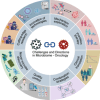
Challenges and future directions of microbiome in the field of precision oncology. This circular diagram illustrates eight key aspects defining the frontier of microbiome‐oncology research: (1) quality control, (2) clinical translation pathways, (3) combination strategies with microbes‐based cancer treatments, (4) personalized therapies based on individual mirobiome profiles, (5) cutting‐edge research developments, (6) new microbial species and their potential application, (7) emerging technological platforms and methodological advances of technologies, and (8) mechanistic insights into biological processes governing microbe‐tumor microenvironment interactions.

References

    1. Human Microbiome Project Consortium . 2012. “Structure, Function and Diversity of the Healthy Human Microbiome.” Nature 486: 207–214. 10.1038/nature11234 - DOI - PMC - PubMed
    1. Kuziel, Gavin A. , Rakoff‐Nahoum Seth. 2022. “The Gut Microbiome.” Current Biology 32: R257–R264. 10.1016/j.cub.2022.02.023 - DOI - PubMed
    1. Virtanen, Seppo , Saqib Schahzad, Kanerva Tinja, Ventin‐Holmberg Rebecka, Nieminen Pekka, Holster Tiina, Kalliala Ilkka, Salonen Anne. 2024. “Metagenome‐Validated Combined Amplicon Sequencing and Text Mining‐Based Annotations for Simultaneous Profiling of Bacteria and Fungi: Vaginal Microbiota and Mycobiota in Healthy Women.” Microbiome 12: 273. 10.1186/s40168-024-01993-9 - DOI - PMC - PubMed
    1. Avershina, Ekaterina , Qureshi Arfa Irej, Winther‐Larsen Hanne C., Rounge Trine B.. 2025. “Challenges in Capturing the Mycobiome From Shotgun Metagenome Data: Lack of Software and Databases.” Microbiome 13: 66. 10.1186/s40168-025-02048-3 - DOI - PMC - PubMed
    1. Rooks, Michelle G. , Garrett Wendy S.. 2016. “Gut Microbiota, Metabolites and Host Immunity.” Nature Reviews Immunology 16: 341–352. 10.1038/nri.2016.42 - DOI - PMC - PubMed

LinkOut - more resources