. 2025 Sep 25;15(9):4043-4053.
doi: 10.62347/BCBI7081. eCollection 2025.

Proteasomal deubiquitinating enzyme USP14/UCHL5 inhibitor bAP15 suppresses endoplasmic reticulum stress-mediated apoptosis and tumor growth in human chondrosarcoma

Affiliations

Proteasomal deubiquitinating enzyme USP14/UCHL5 inhibitor bAP15 suppresses endoplasmic reticulum stress-mediated apoptosis and tumor growth in human chondrosarcoma

Chong-Sun Khoi et al. Am J Cancer Res. .

Abstract

Chondrosarcoma is a malignant bone tumor with limited systemic options. Here, using the small-molecule probe bAP15 to inhibit proteasome-associated deubiquitinating enzymes (DUBs) USP14 and UCHL5, we evaluated the therapeutic concept of DUB inhibition in human chondrosarcoma. Immunohistochemistry showed higher USP14/UCHL5 expression in chondrosarcoma than in normal cartilage controls. In vitro, bAP15 increased Annexin V/PI-positive apoptosis and cleavage of caspase-3/PARP. Under our experimental conditions (low sub-micromolar exposure, 24-48 h), bAP15 led to a predominant accumulation of cells in G1, accompanied by p21 upregulation and reduced PCNA and phospho-histone H3. bAP15 was associated with lower phosphorylation of AKT/ERK and with the induction of ER-stress markers (GRP78, CHOP, IRE1α, caspase-4). In vivo, bAP15 suppressed xenograft growth. Collectively, these data support proteasome-associated DUB inhibition as a potential strategy in chondrosarcoma, with bAP15 serving as a chemical tool to probe this target class.

Keywords: Chondrosarcoma; bAP15; endoplasmic reticulum stress; proteasomal deubiquitinating enzyme.


Conflict of interest statement

None.

Figures

Figure 1
Figure 1
Overexpression of USP14 and UCHL5 in human chondrosarcoma tissues. A. Representative immunohistochemical staining for USP14 and UCHL5 in normal cartilage versus chondrosarcoma tissue. Chondrosarcoma samples show strong positive staining for USP14 and UCHL5 (brown) in tumor cells, whereas normal cartilage exhibits minimal staining. B. Quantification of IHC staining intensity confirming significantly higher USP14 and UCHL5 expression in chondrosarcoma tissues (n=3) compared to normal cartilage (n=3). Data are presented as mean ± SD. P<0.05.
Figure 2
Figure 2
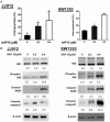
bAP15 induces apoptosis in chondrosarcoma cells in vitro. A. Flow cytometric analysis of apoptosis (Annexin V/PI staining) in JJ012 and SW1353 cells after 48 hours of bAP15 treatment (0, 0.2, 0.4 μM). bAP15 increases the proportion of Annexin V-positive (apoptotic) cells in a dose-dependent manner in both cell lines. Bar graphs indicate the percentage of early and late apoptotic cells at each dose, showing a significant increase in apoptosis compared to vehicle controls. B. Western blot analysis of apoptosis-related proteins in JJ012 and SW1353 cells treated with bAP15 for 48 hours. bAP15 treatment causes cleavage of caspase-3 and PARP and elevates phospho-JNK levels, confirming activation of apoptotic pathways. β-Actin serves as a loading control. Data are representative of three independent experiments. P<0.05 vs control.
Figure 3
Figure 3
bAP15 induces G1 cell cycle arrest in chondrosarcoma cells. A. Cell cycle distribution of JJ012 and SW1353 cells treated with bAP15 (0, 0.2, 0.4 μM) for 48 hours. Propidium iodide DNA content analysis shows an increased G1 population and a corresponding decrease in S and G2/M populations in bAP15-treated cells, indicating G1 phase arrest. Bar graphs depict the percentage of cells in G1, S, and G2/M phases; P<0.05 compared to control for G1 fraction. B. Western blot analysis of cell cycle regulatory proteins in chondrosarcoma cells after bAP15 treatment. bAP15 upregulates p21 and downregulates CDC25C, PCNA, and phospho-histone H3, consistent with G1 arrest. β-Actin is shown as a loading control. These results demonstrate that bAP15 halts cell cycle progression at the G1 phase.
Figure 4
Figure 4
bAP15 suppresses AKT and ERK signaling in chondrosarcoma cells. Western blot analysis of AKT and ERK activation in JJ012 and SW1353 cells following 48-hour bAP15 treatment. bAP15 causes a dose-dependent reduction in phosphorylated AKT (Ser473) and ERK1/2 (Thr202/Tyr204) levels, while total AKT and ERK levels remain unchanged. Densitometric quantification of phospho/total ratios (relative to control) confirms significant suppression of AKT and ERK signaling by bAP15 treatment (P<0.05 vs 0 μM). The loss of AKT/ERK signaling likely contributes to the induction of apoptosis in bAP15-treated cells.
Figure 5
Figure 5
bAP15 triggers endoplasmic reticulum (ER) stress in chondrosarcoma cells. Western blots showing ER stress markers in JJ012 and SW1353 cells after 48 hours of bAP15 exposure. bAP15 upregulates IRE1α, GRP78 (BiP), CHOP, and cleaved caspase-4 compared to untreated controls, indicating activation of the unfolded protein response and ER stress-mediated apoptosis. β-Actin is included as a loading control. Densitometry confirms significant increases in these ER stress markers with bAP15 treatment (P<0.05 vs control).
Figure 6
Figure 6
bAP15 suppresses chondrosarcoma tumor growth in vivo. Nude mice bearing JJ012 or SW1353 chondrosarcoma xenografts were treated with bAP15 (5 mg/kg, intraperitoneally, twice weekly) or vehicle control. A. Tumor growth curves for JJ012 xenografts show that bAP15 significantly inhibits tumor growth compared to vehicle-treated controls. B. Tumor growth curves for SW1353 xenografts similarly demonstrate tumor growth suppression by bAP15. Data are presented as mean tumor volume ± SEM. P<0.01 vs vehicle on the final day of treatment. No significant body weight loss or other toxicity was observed in the bAP15-treated mice, indicating that the treatment was well tolerated.

References

    1. Weinschenk RC, Wang WL, Lewis VO. Chondrosarcoma. J Am Acad Orthop Surg. 2021;29:553–562. - PubMed
    1. Chen JC, Huang C, Lee IN, Wu YP, Tang CH. Amphiregulin enhances cell migration and resistance to doxorubicin in chondrosarcoma cells through the MAPK pathway. Mol Carcinog. 2018;57:1816–1824. - PubMed
    1. Chow WA. Chondrosarcoma: biology, genetics, and epigenetics. F1000Res. 2018;7 F1000 Faculty Rev-1826. - PMC - PubMed
    1. MacDonald IJ, Lin CY, Kuo SJ, Su CM, Tang CH. An update on current and future treatment options for chondrosarcoma. Expert Rev Anticancer Ther. 2019;19:773–786. - PubMed
    1. Speetjens FM, de Jong Y, Gelderblom H, Bovee JV. Molecular oncogenesis of chondrosarcoma: impact for targeted treatment. Curr Opin Oncol. 2016;28:314–322. - PubMed

LinkOut - more resources