. 2025 Oct;55(10):e70079.
doi: 10.1002/eji.70079.

LILRA5 Functions to Induce ROS Production on Innate Immune Cells

Affiliations

LILRA5 Functions to Induce ROS Production on Innate Immune Cells

Zuyi Fu et al. Eur J Immunol. 2025 Oct.

Abstract

Activating immune receptors provides mechanisms for phagocytes to elicit important effector functions that promote the killing of microbes. Leukocyte immunoglobulin-like receptor A5 (LILRA5), an orphan immune receptor expressed by human phagocytes and co-localising with FcRγ, remains poorly characterised. To address this, we developed a highly specific anti-LILRA5 monoclonal antibody that has agonistic properties. We show LILRA5 expression on naïve monocytes and neutrophils, and that ligation of LILRA5 stimulates ROS production. While increased LILRA5 transcripts have been associated with sepsis, we also observed increased levels in patients with systemic infection but without sepsis complications. Ex vivo bacterial infection of whole blood did not alter surface LILRA5 expression, but LPS stimulation changed expression levels, indicating that surface LILRA5 expression is dynamic and likely regulated post-transcriptionally, changing responses to different stimuli or over time. Soluble (s)LILRA5 was enhanced in sera from sepsis patients and in supernatants of monocytes that were LPS-stimulated, indicating that shedding of LILRA5 from cell surfaces or that expression of sLILRA5 isoforms provides a mechanism to regulate surface LILRA5 expression levels. Finally, we show that altered surface LILRA5 expression influences LILRA5-induced ROS production capacity. Thus, LILRA5 is a dynamically regulated activating receptor expressed on phagocytes that stimulates ROS production.

Keywords: LILRA5; activating receptor; infection; reactive oxygen species; sepsis.


Conflict of interest statement

The authors declare no conflicts of interest.

Figures

FIGURE 1
FIGURE 1
A highly specific anti‐LILRA5 antibody with cross‐linking capacity. (A) SDS‐PAGE analysis of rLILRA5‐His. (B) Binding of anti‐LILRA5 P4‐11A mAb to magnetic beads coated with rLILR or control protein, detected using anti‐IgG mAb and flow cytometric analysis. Mean ± SD of n = 3 independent experiments. Paired t‐test. (C) Binding of anti‐LILRA5 clone P4‐11A mAb to U937 cell lines, detected using anti‐IgG mAb and flow cytometric analysis. One representative experiment from n = 3 independent experiments. (D) A schematic of the LILRA5CD3ζ reporter 2B4 T cell line, expressing a surface protein composed of extracellular and transmembrane LILRA5 domains fused to the cytoplasmic tail of CD3ζ. Cross‐linking of CD3 or the fusion LILRA5CD3ζ protein induces ITAM phosphorylation, NFAT activation and GFP expression. (E) Reporter cells stimulated with anti‐CD3 mAb, anti‐LILRA5 P4‐11A mAb, or isotype control. Mean ± SD of n = 4 independent experiments. (F) Detection of rLILRA5‐His by ELISA using anti‐LILRA5 P4‐11A mAb or isotype control. Mean and SD of n = 3 experiments. In all, *p < 0.05 and ****p < 0.0001.
FIGURE 2
FIGURE 2
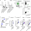
LILRA5 is expressed on human phagocytes and stimulates ROS production. (A) Normalised transcripts per million (nTPM) of LILRA5 in immune cells from the HPA and Monaco datasets. Mean ± SD are shown. (B) Representative example showing the gating strategy used to identify monocytes in human whole blood using anti‐CD14 and anti‐LILRA5 (clone P4‐11A). A first gate was set on physical parameters of SSC‐A vs. FSC‐A, then on SSC‐A and SSC‐H to eliminate doublets, then monocytes and granulocytes were gated on CD14+ and CEACAM8+ (not shown), then on CD14+ events to identify monocytes. (C) Representative flow cytometry staining indicating LILRA5 expression in neutrophils and monocytes from a healthy donor. (D, E) Expression of LILRA5 on human monocytes (D, n = 8) and neutrophils (E, n = 12), determined using anti‐LILRA5 P4‐11A or isotype control. Paired t‐test. (F, G) Reactive oxygen species (ROS) production by PBMCs (F, n = 7) and neutrophils (G, n = 8). One‐way ANOVA of area under the curve (AUC) values, in all, ****p < 0.0001, **p < 0.01, and *p < 0.05.
FIGURE 3
FIGURE 3
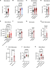
LILRA5 expression during sepsis and systemic infections. LILRA5 expression in whole blood of (A) healthy donors (n = 83), sepsis patients (n = 156; GSE134364). (B) healthy donors (n = 12) or sepsis patients (n = 13), septic shock patients (n = 6; GSE137342). (C) of healthy donors (n = 44) or sepsis patients presenting at emergency rooms (ER) (n = 266), sepsis patients on intensive care units (ICU; n = 82; GSE185263). (D) of healthy donors (n = 8), sepsis patients (n = 20; GSE232753). (E) of healthy donors (n = 40), patients with diagnosed but uncomplicated systemic infection (n = 12), sepsis patients (n = 20), septic shock patients (n = 19; GSE154918). (F) LILRA5 expression in neutrophils from healthy donors (n = 8) or sepsis patients (n = 15; GSE64457). LILRA5 expression in CD14+ monocytes from (G) healthy donors (n = 6), sepsis patients (n = 8; GSE180387), (H) from healthy donors (n = 4), sepsis patients (n = 6; GSE136200), (I) from healthy donors (n = 5), ICU patients with sepsis (n = 4; GSE139913). (J) LILRA5 expression in whole blood from healthy donors (n = 14), patients with bacterial infections (n = 24), and patients with viral infections (n = 28; GSE72810). (K) LILRA5 expression in whole blood from healthy donors (n = 43), patients with E. coli infection (n = 32), patients with S. aureus infection (n = 19; GSE33341). In all panels, mean ± SD are shown. Statistics tested by limma, where adjusted ****p < 0.0001, ***p < 0.001, and *p <0.05.
FIGURE 4
FIGURE 4
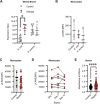
Surface LILRA5 expression does not increase during infection and inflammation. (A) Whole blood LILRA5 expression after ex vivo infection with E. coli (n = 5, control n = 8) or S. aureus (n = 5, control n = 8; GSE65088). Mean ± SD are shown. Statistics tested by limma. (B) Expression of LILRA5 on human monocytes after ex vivo infection of whole blood by E. coli or S. aureus, from n = 3 independent experiments. (C) Surface LILRA5 expression on human monocytes from healthy donors (n = 8) and sepsis patients (n = 26), upon hospital admission. Mean ± SD are shown. (D) Surface LILRA5 expression on human monocytes from sepsis patients (n = 8) at the indicated days since hospital admission. (E) Comparison of sLILRA5 in serum from sepsis patients (n = 128) or healthy donors (n = 60). Mean ± SD are shown. Student t‐test. In all, ****p < 0.0001, ***p < 0.001. **p < 0.01, and *p < 0.05.
FIGURE 5
FIGURE 5
LPS activation reduces LILRA5‐dependent ROS production. (A) LILRA5 expression by monocytes cultured ± LPS for 18 h (GSE147310). Data from n = 5 donors. Statistics tested by limma. (B) Surface LILRA5 expression on human monocytes from healthy donors (n = 3). Student t‐test. (C) sLILRA5 in culture supernatants from PBMCs from healthy donors (n = 4), after 18 h culture ± LPS. Student t‐test. (D, E) Production of ROS by PBMCs in response to LILRA5 ± LPS stimulation. Data from n = 5 independent donors. Raw ROS production values are shown in (D). Student t‐test. The ROS production induced by anti‐LILRA5 P4‐11A relative to IgG1 is compared through area under the curve (AUC), as shown in (E). In all, **< 0.01, *p < 0.05.

References

    1. Dale D. C., Boxer L., and Liles W. C., “The Phagocytes: Neutrophils and Monocytes,” Blood 112 (2008): 935–945. - PubMed
    1. Underhill D. M., Rossnagle E., Lowell C. A., and Simmons R. M., “Dectin‐1 Activates Syk Tyrosine Kinase in a Dynamic Subset of Macrophages for Reactive Oxygen Production,” Blood 106 (2005): 2543–2550, 10.1182/blood-2005-03-1239. - DOI - PMC - PubMed
    1. Ravetch J. V. and Bolland S., “IgG Fc Receptors,” Annual Review of Immunology 19 (2001): 275–290, 10.1146/annurev.immunol.19.1.275. - DOI - PubMed
    1. Huizinga T. W., van Kemenade F., Koenderman L., et al., “The 40‐kDa Fc Gamma Receptor (FcRII) on Human Neutrophils Is Essential for the IgG‐Induced Respiratory Burst and IgG‐induced Phagocytosis,” Journal of Immunology 142 (1989): 2365–2369, 10.4049/jimmunol.142.7.2365. - DOI - PubMed
    1. Lee W. B., Kang J. S., Yan J. J., Lee M. S., et al., “Neutrophils Promote Mycobacterial Trehalose Dimycolate‐Induced Lung Inflammation via the Mincle Pathway,” Plos Pathogens 8 (2012): e1002614, 10.1371/journal.ppat.1002614. - DOI - PMC - PubMed

MeSH terms

LinkOut - more resources