. 2025 Oct 21;15(1):36627.
doi: 10.1038/s41598-025-20542-4.

Implantation of a vascular access button in mice

Affiliations

Implantation of a vascular access button in mice

Suguru Yamauchi et al. Sci Rep. .

Abstract

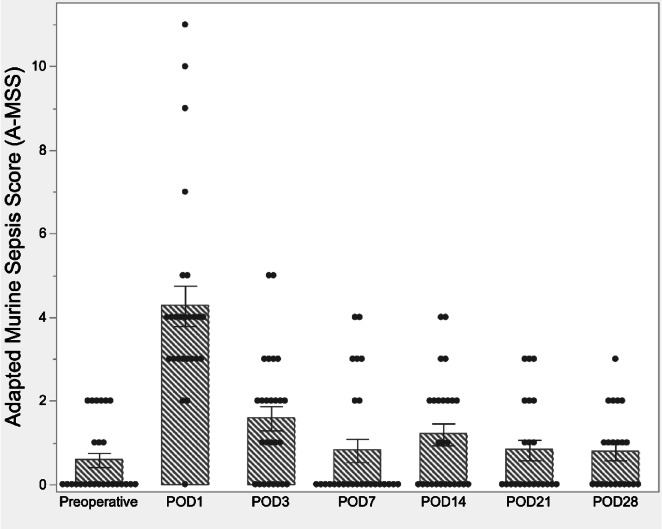
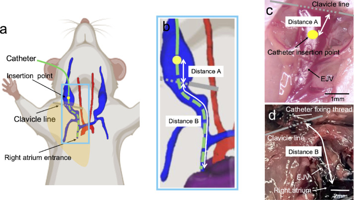
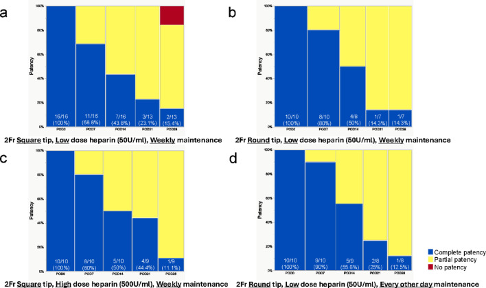
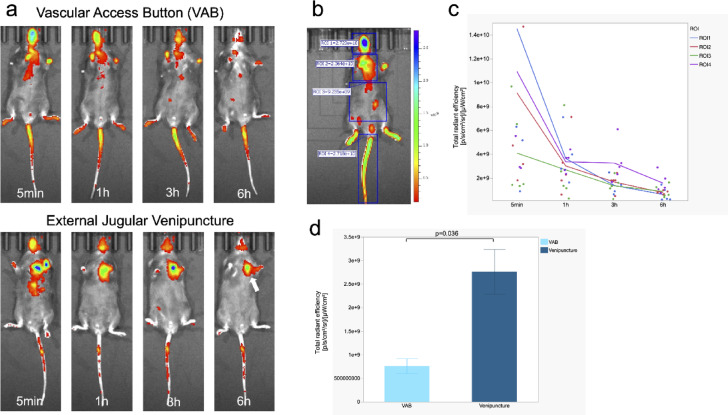
Vascular access presents unique challenges in experimental mice due to their small size and anatomical constraints. Achieving reliable vascular access is crucial for optimizing experimental outcomes, especially in protocols requiring serial blood sampling or repeated intravascular therapy. Although vascular access buttons (VABs) offer significant advantages, their widespread adoption has been limited by technical challenges and a lack of comprehensive validation regarding their safety, feasibility, and long-term management. To address these gaps, we conducted a comprehensive evaluation of VAB implantation in mice. The technical success rate was 90.2%, and the 28-day survival rate was 80.4%. Optimal catheter insertion lengths determined by intraoperative and autopsy findings, and computed tomography were 9.5 ± 0.6 mm (20-25 g), 10.1 ± 0.8 mm (25-30 g), and 11.2 ± 0.5 mm (> 30 g), respectively. Catheter patency was analyzed by stratifying the cohort into groups based on physical parameters such as catheter tip geometry, heparin concentration in the maintenance solution, and frequency of catheter maintenance procedures. Although approximately half of the mice lost complete catheter patency by day 14, the majority maintained partial patency at day 28 in all experimental groups except for two cases of complete occlusion in the 2 Fr square tip, low-dose heparin, weekly maintenance group. The evaluation of biodistribution and clearance with indocyanine green indicated that VAB administration may have advantages over conventional venipuncture. These standardized methodologies can provide a framework for diverse biomedical research applications, enhancing both the efficiency and reproducibility of studies requiring reliable vascular access.

Keywords: External jugular vein; Vascular access; Vascular access button; Vascular catheter.


Conflict of interest statement

Declarations. Competing interests: The authors declare no competing interests.

Figures

Fig. 1
Fig. 1
Experimental design. Flowchart for all analyses of this study. VAB, vascular access button; CT, contrast-enhanced computed tomography.
Fig. 2
Fig. 2
Adapted murine sepsis score (A-MSS) over the course of the postoperative period. The A-MSS score, a metric used to assess postoperative recovery in mice, typically reached its peak value on postoperative day 1 (POD1). Subsequently, the score demonstrated a gradual improvement, with most subjects returning to near-baseline values by POD7.
Fig. 3
Fig. 3
Schematic and image of catheter length. (a) An overall view shows catheter insertion through the external jugular vein. Blue indicates the external jugular and subclavian veins; red, the aortic arch and carotid artery; and purple, the heart. Green denotes the catheter; yellow indicates the point of insertion of the catheter into the external jugular vein. (b) Close-up view of Fig. 1a. Distance A is defined as the distance from the external jugular vein insertion point of the catheter to the clavicular line. Distance B is defined as the distance from the clavicular line to the superior vena cava-right atrial junction. (c) Full view of the external jugular vein in surgery. Distance A is determined intraoperatively. (d) Full view of the external jugular vein into which the catheter was inserted at autopsy. The sternum and ribs have been removed. Distance B was measured manually at autopsy. Distance A plus Distance B is the optimal catheter insertion length (catheter tip at the junction of the superior vena cava and right atrium) into the external jugular vein. EJV, external jugular vein.
Fig. 4
Fig. 4
Comparison of different catheter insertion lengths in living mice. Imaging after catheter insertion obtained from an additional cohort of living mice (n = 5) allowed visualization and confirmation of optimal catheter tip location. Blue indicates the external jugular and subclavian veins; red, the aortic arch and carotid artery; and purple, the heart. A white high-intensity catheter filled with a contrast agent can be recognized as a curved material connected to the VAB. (a) 3D reconstruction demonstrating anterior view of a VAB with 9-mm catheter placement to the cardiovascular system of a 21 g mouse. (b) Oblique view of a 3D rendering of a 9 mm long VAB with catheter placement to the cardiovascular system of a 21 g mouse (c) Confirmation of the catheter tip on contrast-enhanced CT cross-sectional image. White arrow; the tip of the catheter located in the superior vena cava just before the right atrial entrance. (d) Anterior view of a 3D rendering of a 10 mm long VAB with catheter placement to the cardiovascular system of a 26 g mouse. (e) Anterior view of a 3D rendering of an 11 mm long VAB with a catheter implanted in a 30 g mouse. BW, body weight; CL, catheter length.
Fig. 5
Fig. 5
Catheter patency over time with different management methods. (a) The catheter tip geometry is square, heparinized saline used for flushing is a low dose (50U/ml), and catheter maintenance is weekly. (b) The catheter tip geometry is round, heparinized saline used for flushing is low dose (50U/ml), and catheter maintenance is weekly. (c) The catheter tip geometry is square, heparinized saline used for flushing is a high dose (500U/ml), and catheter maintenance is weekly. (d) The catheter tip geometry is round, heparinized saline used for flushing is low dose (50U/ml), and catheter maintenance is every other day. The numbers shown in the figure represent the percentage of complete patency.
Fig. 6
Fig. 6
Catheter patency over time with 3Fr catheter. 3Fr diameter, the catheter tip geometry is square, heparinized saline used for flushing is low dose (50U/ml), and catheter maintenance is every other day. The numbers shown in the figure represent the percentage of complete patency.
Fig. 7
Fig. 7
Biodistribution and clearance of the ICG with IV Injection via VAB. (a) Comparison of VAB vs. direct external jugular venipuncture over time. In both routes of administration, ICG is distributed throughout the body immediately after administration and is excreted in approximately six hours. Venipuncture mice still show relatively high ICG fluorescence at 6 h post-evaluation (White arrow). (b) The head, chest, abdomen, and tail were defined as regions of interest (ROI)1, ROI2, ROI3, and ROI4, respectively, and total radiant efficiency was calculated. (c) Trends over time in the total radiant efficiency of each ROI in mice administered ICG via VAB. The metabolic and excretory processes of ICG administered via VAB are represented by the quantitative decrease in fluorescence intensity in all ROIs. (d) Comparison of total radiant efficiency in ROI2 area at 6 h in VAB and venipuncture. Venipuncture mice have significantly higher fluorescence intensity (p = 0.036, Mann–Whitney U-test).

References

    1. Waterston, R. H. et al. Initial sequencing and comparative analysis of the mouse genome. Nature420, 520–562. 10.1038/nature01262 (2002). - PubMed
    1. Breschi, A., Gingeras, T. R. & Guigó, R. Comparative transcriptomics in human and mouse. Nat. Rev. Genet.18, 425–440. 10.1038/nrg.2017.19 (2017). - PMC - PubMed
    1. Slosky, L. M. et al. Establishment of multi-stage intravenous self-administration paradigms in mice. Sci. Rep.12, 21422. 10.1038/s41598-022-24740-2 (2022). - PMC - PubMed
    1. Burvin, R., Zloczower, M. & Karnieli, E. Double-vein jugular/inferior vena cava clamp technique for long-term in vivo studies in rats. Physiol. Behav.63, 511–515. 10.1016/s0031-9384(97)00486-1 (1998). - PubMed
    1. Noble, F., Coric, P., Turcaud, S., Fournié-Zaluski, M. C. & Roques, B. P. Assessment of physical dependence after continuous perfusion into the rat jugular vein of the mixed inhibitor of enkephalin-degrading enzymes, RB 101. Eur. J. Pharmacol.253, 283–287. 10.1016/0014-2999(94)90203-8 (1994). - PubMed

LinkOut - more resources