Review
. 2025 Oct;55(11):2400-2416.
doi: 10.1007/s00247-025-06431-y. Epub 2025 Oct 22.

Fetal cardiac masses

Affiliations
Review

Fetal cardiac masses

Bhagyashree Rathore et al. Pediatr Radiol. 2025 Oct.

Abstract

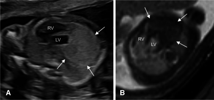
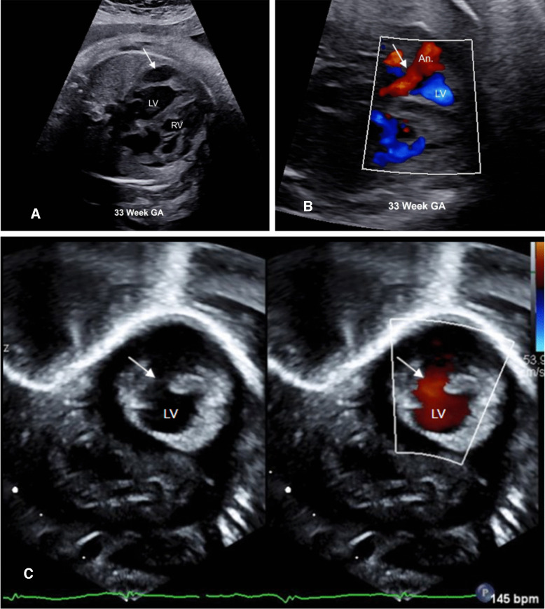
Although rare in the pediatric population, cardiac tumors represent a significant portion of prenatally detected masses, second only to sacrococcygeal teratomas. Fetal cardiac masses are predominantly benign, with rhabdomyoma being the most common fetal cardiac mass in the setting of tuberous sclerosis. Multiple lesions are pathognomonic for rhabdomyomas and prompt evaluation for tuberous sclerosis. Fibroma, teratoma, and hemangioma are other consistently detected cardiac tumors. In this article, we review the epidemiology, imaging, treatment, and outcomes of these fetal cardiac masses, emphasizing the role of imaging in surveillance, identifying complications, refining the differential diagnosis, and recognizing potential diagnostic pitfalls.

Keywords: Fetus; Fibroma; Heart neoplasms; Hemangioma; Rhabdomyoma; Teratoma.


Conflict of interest statement

Declarations. Conflicts of interest: None

Figures

Fig. 1
Fig. 1
A 33-week gestational age (GA) fetus with confirmed TSC2 gene mutation. A Axial T2-weighted image shows multiple T2 hyperintense ovoid lesions (arrows) involving the myocardium and inter-ventricular septum. B Four-chamber echocardiography image demonstrating multiple ovoid hyperechoic masses (arrows) in the same location as the MR. C Axial balanced steady state free precession (bSSFP) brain image showing a hypointense subependymal nodule (arrow) along the right lateral ventricle. The patient received mTOR inhibitors. D Axial T2-weighted image of the brain at 3 years of age showing multiple cortical, subcortical lesions/tubers (arrows), and subependymal nodules (arrowhead) typical of tuberous sclerosis. Postnatal echocardiogram in an apical 4-chamber view confirmed the presence of rhabdomyomas (arrow) in the same locations identified prenatally (E), but over time they reduced in size, with the most recent echocardiogram performed at 5 years of age showing no intra-cardiac tumors (F). LV, left ventricle; RV, right ventricle; LA, left atrium; RA, right atrium
Fig. 2
Fig. 2
A fetal cardiac mass detected in a 20-week gestational age pregnancy. A Horizontal view echocardiogram demonstrating a large, well-defined hyperechoic mass at the cardiac apex (arrows). B Axial T2-weighted image shows a large solitary T2 hypointense mass (arrows). No additional lesions were seen. Imaging of fetal brain and kidneys was unremarkable. The differential considerations were fibroma or rhabdomyoma. Unfortunately, the pregnancy resulted in fetal demise, with genetic testing positive for tuberous sclerosis and an autopsy confirming a rhabdomyoma. LV, left ventricle; R right ventricle
Fig. 3
Fig. 3
A Sagittal and (B) axial T2-weighted images from fetal MRI performed at 32 weeks gestational age demonstrating a complex cystic mass (arrows) centered in the right mediastinum with mass effect on the right atrium. C Axial horizontal view fetal echocardiogram at 28 weeks gestational age showing a heterogeneous echogenic mass (arrows) in the right chest adjacent to the right atrium and right ventricle. D Follow-up axial horizontal view fetal echocardiogram at 34 weeks gestational age shows increased size of the mass (arrows) with increased mass effect on the right atrium. The patient was delivered via C-section at 34 weeks 1 day due to the worsening hydrops and underwent immediate resection of the mass. Histopathology was consistent with congenital immature teratoma with no malignant components. LA, left atrium; LV, left ventricle; RV, right ventricle, GA, gestational age
Fig. 4
Fig. 4
A Axial T2 and B balanced steady state free precession (bSSFP) images in a 29-week gestational age fetus demonstrating a well-defined mass (arrows) in the right ventricular wall, confirmed fibroma postnatally. RV, right ventricle; GA, gestational age
Fig. 5
Fig. 5
A Axial T2-weighted and (B) balanced steady state free precession images at 32 weeks gestational age showing a T2 hyperintense, balanced steady state free precession iso-hypointense mass (arrow) associated with the right atrium. Follow-up axial (C) T2 and (D) Balanced steady state free precession images at 38 weeks gestational age show increased size of the lesion (arrows). It is also more heterogeneous on T2-weighted images and is causing mass effect on the right atrium. E The sagittal fast spoiled gradient echo image does not demonstrate any intra-lesional hyperintensity to suggest fat or hemorrhage. F Axial 4-chamber view at 32 weeks gestational age demonstrates a homogeneous hyperechoic lesion (arrow) effacing the right atrium. G Postnatal parasternal long axis view echocardiogram angled toward the right ventricle at day of life 1 demonstrates the echogenic mass (arrow) with no appreciable intra-lesional flow on color Doppler on split screen view. The lesion was resected, and histopathology was consistent with a rapidly involuting congenital hemangioma. RA, right atrium; LA, left atrium; RV, right ventricle; LV, left ventricle; GA, gestational age
Fig. 6
Fig. 6
A Axial T2-weighted image in a 20-week gestation pregnancy showing a well-defined T2 hyperintense lesion (arrow) along the right cardiac border. B An axial fetal echocardiogram image in split screen view centered near the entrance of the inferior vena cava to the right atrium showing a well-defined anechoic cystic lesion (arrow) with no vascularity on color Doppler along the right cardiac border. GA, gestational age
Fig. 7
Fig. 7
A Echocardiogram in a 33-week gestation fetus referred for evaluation of arrhythmia demonstrating hypoechoic lesion in the left ventricular wall (arrow). B Color Doppler image showing gap in the left ventricular wall (arrow) with flow into the aneurysm (An). C Postnatal echocardiography from a subcostal sagittal view confirmed the diagnosis of ventricular aneurysm. The LV to aneurysmal connection (arrow) is shown on split screen image. LV, left ventricle; RV, right ventricle; An, aneurysm

References

    1. Kwasniewicz P, Wieczorek-Pastusiak J, Romaniuk-Doroszewska A, Bekiesinska-Figatowska M (2023) Congenital tumors-magnetic resonance imaging findings with focus on rare tumors. Cancers (Basel). 10.3390/cancers16010043 - PMC - PubMed
    1. Camargo FM, Brizot MdL, Francisco RPV, de Carvalho WB, Ikari NM, Peres SV, Lopes MAB, Lopes LM (2024) Resultados perinatais e seguimento em Longo prazo de tumores cardíacos fetais: Estudo de coorte histórica de 30 anos. Arq Bras Cardiol. Sociedade Brasileira de Cardiologia 121(1):e20220469 - PMC - PubMed
    1. Van der Hauwaert LG (1971) Cardiac tumours in infancy and childhood. Br Heart J 33:125–132 - PMC - PubMed
    1. Hornberger LK, McBrien A (2018) Fetal cardiac tumors. In: Yagel S, Silverman NH, Gembruch U (eds) Fetal cardiology: Embryology, genetics, physiology, echocardiographic evaluation, diagnosis, and perinatal management of cardiac diseases, 3rd edn. Philadelphia: CRC Press
    1. Kogon B, Shehata B, Katzenstein H, Samai C, Mahle W, Maher K, Olson T (2011) Primary congenital infantile fibrosarcoma of the heart: the first confirmed case. Ann Thorac Surg 91:1276–1280 - PubMed

LinkOut - more resources